
Dr. Adam Wexler, Post-Doctoral Researcher
Arie Zwijnenburg Laboratory for Advanced Microscopy and Optical Metrology,
Wetsus – European Centre of Excellence for Sustainable Water Technology, Leeuwarden, Netherlands
Background
Dr. Wexler’s research interest is to bring the power of photonics to the field of sustainable water technology. Currently he, along with collaborators at the University of Twente, are focused on preventing outbreaks of waterborne viruses by understanding and improving water filtration technologies.
The virus particles investigated are small, roughly 20-30 nm in size, and can sometimes pass through water filtration systems and remain infectious. There is currently no practical way to investigate the effectiveness of these filtration devices in real time.
Dr. Wexler is working towards building a microscope that permits tracking of these particles in real time to detect viable viruses in the water and assess the efficiency of these filtration processes. The ultimate aim is to couple the real-time imaging to automated imaging algorithms that can detect problems in the water and alert water production managers before an outbreak begins.
Challenge
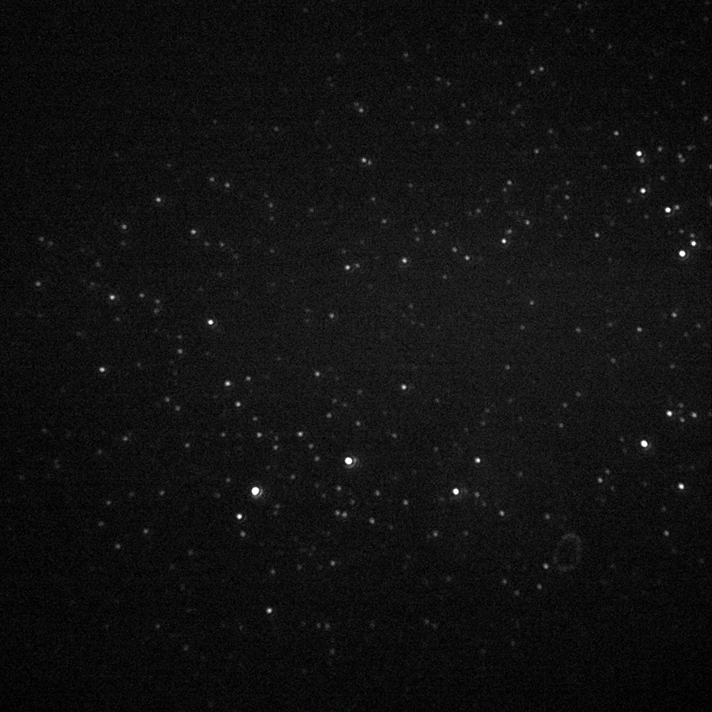
The primary challenge is the low fluorophore labelling densities, Dr. Wexler explains that they try not to modify the viruses too much so there are very few fluorophores (<40) per virus.
Dr. Wexler and his colleagues realized that when there are 40 fluorophores on a virus capsid they exhibit stable fluorescence but when the viruses are fragmented they blink. This allows differentiation between whole and broken virus particles. The combination of low fluorophore density, rapid blinking, and flow means that low noise, camera speed, and sensitivity are important.
Dr. Wexler looked at several sCMOS cameras and told us that one of the biggest factors for them was the quantum efficiency (QE). He sometimes uses far-red dyes so a high QE at these wavelengths is of additional importance.
The increase in QE was huge, and really helped with our application.
Solution
Dr. Wexler is now using the Teledyne Photometrics Prime BSI back-illuminated sCMOS camera on his widefield microscope to image these virus particles in real-time under flow at 40× magnification. He shared, “The increase in QE was huge, and really helped with our application.”
Dr. Wexler also mentioned that the larger field of view was a welcome improvement because they are tracking and counting particles, so the increased throughput really improves the quality of statistics from the population.
Dr. Wexler concluded, “So far the camera performs well and has a lot of potential for our future work.” They also hope that the high speed will be useful for quantification measurements using FRET to distinguish between whole and fragmented viral particles.
