
Prof. Jiulin Du
Dr. Jianan Liu
Dr. Rongwei Zhang
Institute of Neuroscience, Shanghai Research Center for Brain Science and Brain-Inspired Intelligence, Chinese Academy of Sciences (CAS), Shanghai
Background
Prof. Jiulin Du’s group is engaged in understanding the mechanisms of how multiple sources of sensory information are integrated in our central nervous system in order to execute behaviors. They investigate neuronal activity through in vivo electrophysiological recording and optical imaging on the model organism zebrafish. However, such a recording strategy is invasive and difficult to scale up for high throughput recording, while other options such as calcium imaging ignore the subthreshold membrane potential fluctuations which also play an essential role in the development of neuronal computation. Because of this, they hope to develop a novel near-infrared (NIR)-excited voltage nanosensor that can directly monitor the changes in membrane potential.
Most optical voltage sensors need to be excited by high-intensity visible light, these wavelengths of light suffer from distortion and scattering in deep tissues, and prevent long-term imaging due to photobleaching. NIR excitation has deeper penetration and lower phototoxicity, permitting long-term recording of membrane potential fluctuations of neurons from deep brain areas.
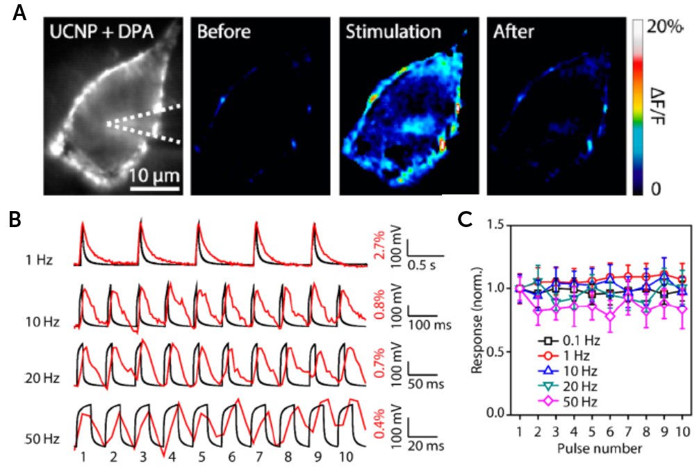
A: Pseudocolor images showing luminescence change of UCNP-labeled HEK293 cells before, during, and after 100 mV depolarization. B/C: Examples and summary of UCNP luminescence changes (red line) in response to repetitive depolarization (black line) of different frequencies (0.1, 1, 10,20, and 50 Hz).
Challenge
The nanosensor is constructed by engineering Förster resonance energy transfer (FRET) efficiency between the outer membrane-anchored upconversion nanoparticle (UCNP), and the membrane-embedded dipicrylamine (DPA). When the neuron is at resting state, FRET is occurring and the luminescence of UCNPs are absorbed by DPA. When the neuron is depolarized, FRET stops and the luminescence of UCNPs is increased. As a result, the emission intensity of the nanosensor can report changes in membrane potential.
The FRET signal is dim and shows a small response to voltage changes, so for this application, Prof. Du’s group needed a camera with high sensitivity to capture as many photons as possible. In addition, since neuronal spikes only last a few milliseconds, a high-speed camera with a fast scanning rate is necessary to detect the membrane potential changes.
Compared to other EMCCD and sCMOS cameras, the Prime 95B can detect very dim signals, achieving a high signal-to-noise ratio and fast imaging rate, giving good spatiotemporal resolution.
Solution
After comparing different EMCCD and sCMOS cameras, Prof Du’s group chose the Teledyne Photometrics Prime 95B sCMOS camera to detect their FRET signals.
Dr. Rongwei Zhang, an associate researcher from the Du lab, told us: “Prime 95B has a very high quantum efficiency, which is really helpful for our voltage nanosensor imaging… We used Prime 95B to detect membrane potential-relevant fluorescent signals for the use of our voltage nanoparticle sensors in cultured cells and zebrafish larvae. Compared to other EMCCD and sCMOS cameras, [the] Prime 95B can detect very dim signals, achieving a high signal-to-noise ratio and fast imaging rate, giving good spatiotemporal resolution.”
