Dr. Asaph Zylbertal, Dr. Issac Bianco
Department of Neuroscience, Physiology & Pharmacology, University College London, UK
Background
The Bianco Lab at UCL aims to understand the logic of brain circuits that control behavior. Using two‑photon and light-sheet microscopy with small, optically transparent larval zebrafish expressing genetically encoded calcium indicators (GECIs), researchers such as Dr. Asaph Zylbertal record activity at single-cell resolution throughout the brain.
Dr. Zylbertal explained more about his work, “I am using our light-sheet microscope to look at tens of thousands of neurons expressing GECIs across the whole zebrafish brain, to see activity and estimate what each individual neuron in the fish brain is doing at any given time.”
“After acquiring the raw data, we use a segmentation algorithm to find neurons in each imaging plane, we then deconvolve the calcium signal from these neurons to estimate spikes, this gives us a representation of activity across the brain.”
While some light sheet experiments look at the anatomy of the zebrafish, the aim of Dr. Zylbertal is to observe functional activity within the zebrafish brain.

Figure 1: Video stack of the brain of a zebrafish larva, taken with the Kinetix sCMOS. Each image is of a different z-plane through the sample, which is expressing the GCaMP6f calcium indicator under the elavl3 promotor (elavl:GCaMP6f, Wolf et al. 2017). Dorsal view, anterior side is up, the view is approx. 420 x 710 x 150 μm3.
Challenge
Calcium imaging requires high speeds in order to capture the dynamic signaling between neurons. However, to monitor populations of cells throughout the brain, it is also necessary to capture large imaging volumes. Thus, the detector needs to be capable of both high speed and a large field of view (FOV) for this high‑throughput application.
Dr. Zylbertal told us about the experimental challenges, “When we are using fast indicators, we would like to be able to image as many volumes as possible over time, all with high resolution so we can integrate lots of pixels in order to get a higher signal to noise.”
This application needs a combination of speed and sensitivity, all across a large FOV.
The Kinetix allows us to image a large field of view at high speeds and with high sensitivity, which is ideal for our research.
Solution
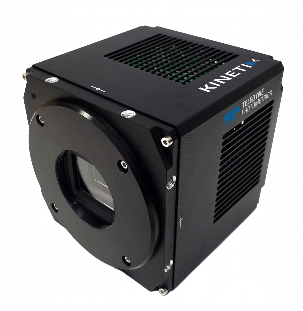
The Kinetix is an ideal solution for this application, featuring extremely high speeds (500 fps across the full frame), high sensitivity (<1 e- read noise and 95% QE signal collection), and a huge FOV (29 mm diagonal), all in one camera.
The Kinetix Speed Mode operates at 500 Hz across the full 10 megapixel sensor, enabling the Kinetix to capture rapid calcium signals from neurons in real-time. Combining this with the high sensitivity and large FOV allows for volumetric functional imaging across the whole zebrafish brain while retaining high resolution to resolve individual neurons.
Dr. Zylbertal outlined his experience with the Kinetix, “The advanced triggering is very important. We synchronize the camera with the light sheet and use pseudo-global shutter”
“With the Kinetix, we are imaging at a larger FOV and a higher speed than previously, which is what we need for this application. Experiments can last for hours and we want to react to the images as they come. With the Kinetix, we have the higher signal to noise ratio needed to extract activity from the neurons.”

Reference
Wolf, S., Dubreuil, A.M., Bertoni, T. et al. (2017) Sensorimotor computation underlying phototaxis in
zebrafish. Nature Communications 8, 651. https://doi.org/10.1038/s41467-017-00310-3
