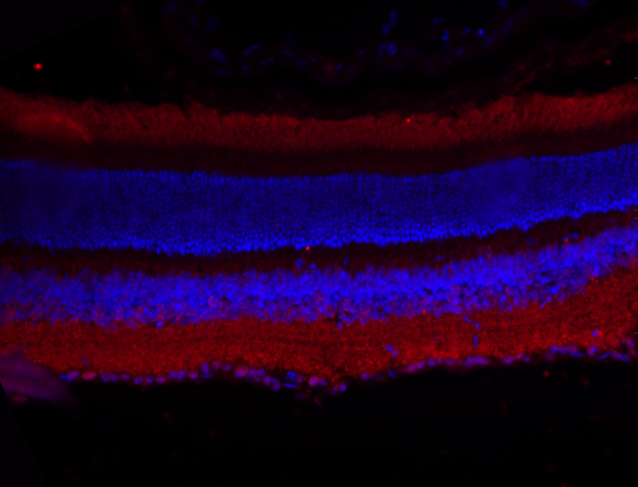
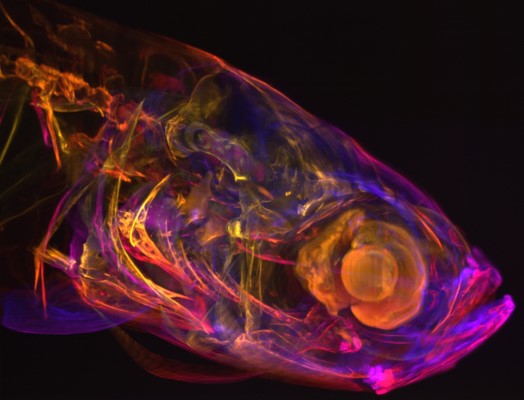
Fluorescence Microscopy
In microscopy, it is vital to have some form of contrast or stain that gives areas of the sample color and makes them possible to image. In addition, it’s often desirable to image just some of the structures inside a cell, such as the nucleus or mitochondria. Fluorescence imaging allows for both of these things.
Fluorescent molecules (known as fluorophores) are used to label samples, and fluorophores are available that emit light in virtually any color. By localizing these fluorophores to the area of interest a clear image of any part of a cell can be taken, making fluorescence microscopy a powerful tool for life sciences.
There are multiple techniques within fluorescence microscopy, each with different equipment requirements including scientific cameras.
Fixed Sample And Documentation Imaging
Capturing, documenting and analyzing color and monochrome images requires a scientific camera capable of producing live images at video rates with high resolution and sensitivity for the highest, publication-ready quality.
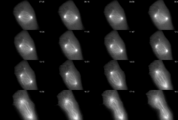
Live Cell Imaging
Live cell imaging is the observation of dynamic processes in cells, tissues, or whole organisms as they happen. Compared to fixed cells, live cells provide more information about the changes that occur in the cell during processes necessary for life. This includes everything from cell division to cell migration, movements and transformations of organelles and calcium imaging.

Spinning Disk Confocal Imaging
Confocal microscopy uses optical sectioning to take multiple, thin, 2-dimensional slices of a sample to construct a 3-dimensional model from them. This is made possible through the addition of a pinhole into the same focal plane as the sample to block out-of-focus light.
Spinning disk confocal microscopy increases the speed of this technique by using multiple pinholes etched into an opaque disk which, when spun, scans the pinholes across the entire image.
Light Sheet Microscopy Imaging
Light sheet microscopy enables scientists to overcome two major problems in modern microscopy. Namely, to image biological samples for much longer under physiologically relevant conditions than with conventional microscopy techniques and to image samples of considerable size in a more reasonable and relevant time frame.
Single Molecule Imaging
Single-molecule fluorescence microscopy represents a subset of fluorescence microscopy that uses fluorescent tags to detect and analyze individual single molecules. This allows the activity of single molecules to be visualized with high signal-to-noise without disturbing the physiological conditions of the biological system.
TIRF Imaging
Total internal reflection fluorescence microscopy (TIRF) makes use of specific optics to produce illumination light only at the 50-100 nm range at the interface of the slide, massively reducing out of focus light and improving the ability to detect fluorescent molecules. Because of its low light intensity and high spatial resolution, it is a key technique in live-cell imaging.

Super Resolution Imaging
Many molecules and structures of interest require a higher degree of resolution than standard microscopy techniques, so it was necessary to develop a technique to break the diffraction limit of light to see them. There are now a significant number of super-resolution techniques such as localization based techniques (PALM/STORM), structural techniques (SIM/iSIM) and post-processing techniques (SRRF/SOFI).
Neuroscience And Electrophysiology Imaging
Advanced imaging methods in neuroscience allow for imaging deep into brain tissue with high spatial and temporal resolution. It is even possible, using advanced optogenetic methods, to optically interrogate cells to discover more about their function.