
Neuroscience
Advanced imaging methods in neuroscience permit us to image deep into brain tissue with high spatial and temporal resolution. It is even possible, using advanced optogenetic methods, to optically interrogate cells to discover more about their function.
Electrophysiology is a method that allows us to study the function of electrically active cells, such as neurons. Research on the physiology of neurons and the nervous system is often accomplished at the cellular level using microscopy techniques or further techniques such as patch clamping.
For a quick overview of neuroscience, take a look at our short article: What is Neuroscience?


Electrophysiology
In many electrophysiology research applications, probes are used to measure the electrical activity of individual cells, tissues, and whole specimens.
Measuring the activity of cells at various membrane potentials can give valuable information about ion transport mechanisms and cellular communication. Varying ionic strength and membrane potential on cell populations can be used to study contractile movements of muscle cells, and diseases affecting the normal propagation of impulses. Using various stimulation techniques along with a selection of quality equipment and setup design can give rise to a wealth of applications in electrophysiology.
Calcium Imaging
Calcium (Ca2+) is one of the most relevant ions in the body as it is essential for e.g. the accurate timing and function of interneuronal communication or cardiomyocytes.
In a physiological system, intracellular and extracellular Ca2+ concentrations are usually not in equilibrium and the concentration gradient is maintained by the cell with a very complex system of ion channels and transporters.
The importance of Ca2+ in physiological systems makes it a very active area of research and it is crucial to be able to monitor Ca2+ dynamics in a reliable manner.


Intrinsic Signal Optical Imaging
Intrinsic signal optical imaging (ISOI) is a technique used to map dynamics in single cells, brain slices and even – most importantly – entire mammalian brains. It is label-free, minimally invasive and gives the researcher access to the basic physiology of a functioning brain.
All of these benefits come without having to compromise on spatial and temporal resolution which is otherwise only provided by regular imaging techniques that rely on labels and markers. As the use of the latter techniques is undesired in humans, ISOI can be used to confirm observations made with optical imaging techniques in other mammals.
Optogenetics
Optogenetics combines optical and genetic techniques to allow direct control of electrical and biochemical events, such as the firing or suppression of neurons, triggered by direct light stimulation.
This is achieved by introducing proteins with light-activated channels, called Opsins, into the cells of interest using targeted genetic techniques. Once in place, these channels allow manipulation of neurons on timescales of milliseconds rather than months. The result is a technique that can be used to both study the role of neurons and control the behavior of an organism, either in vivo or in vitro.


Voltage Imaging
Voltage imaging involves using fluorescent voltage indicators in order to directly image voltage changes in neurons. There are constant advancements in voltage imaging, with voltage indicators ranging from small molecule voltage-sensitive dyes to genetically-encoded voltage indicators, while remaining non-destructive.
Modern voltage imaging can display cell signals with subcellular spatial resolution and fast temporal resolution, enough to match neuronal cell signaling. In terms of speed, voltage image is a very fast technique and has been used in many experiments on live cells and large model organisms.
The Retiga ELECTRO for Electrophysiology
Electrophysiology applications require the user to have intimate knowledge and control of every component of the system.
The selection of a camera for electrophysiology is of great importance to properly sample positioning and data collection. The Retiga ELECTRO offers electrophysiologists an ideal solution to imaging challenges commonly present with other cameras.
Advanced Cameras for Electrophysiology
There are some cases where electrophysiology imaging requirements go beyond the Retiga ELECTRO.
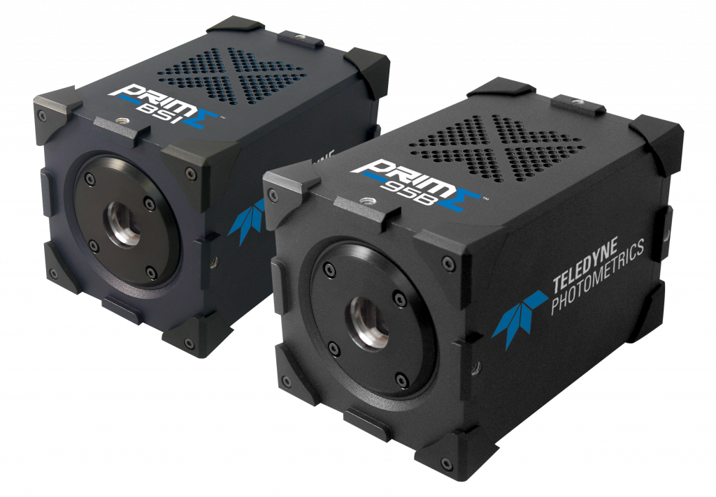
For this reason, Teledyne Photometrics recommends two other camera types that may better suit these applications; the Iris series of cameras for high resolution, large field of view imaging and the Prime series of cameras for ultimate sensitivity, speed and contrast.