
Alexandros Fragkopoulos, PhD
Oliver Bäumchen, PhD, Group Leader
Max Planck Institute for Dynamics and Self Organization
Physics of Soft and Living Matter, Bäumchen Lab, Göttingen
Background
The Bäumchen lab is researching the physics underlying interfaces of soft and biological matter. In particular, they want to understand how those interfaces can alter the dynamics of soft and living matter. To achieve this, a multitude of techniques including micro- and nanofluidics, lab-on-a-chip technologies and force spectroscopy methods are employed. One of the lab’s model systems is the microalgae Chlamydomonas reinhardtii, for which the lab is studying flagella-mediated cell adhesion and motility, at interfaces which can be controlled by light.
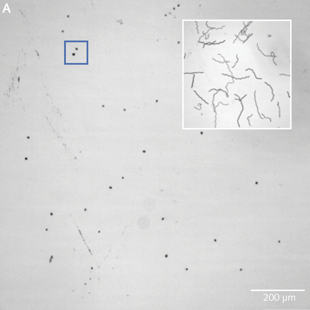
A) shows the entire full field of view. The insert (top right) is a minimum intensity projection showing the trajectories of individuals. B) Every 10th frame from this movie and C) a series of consecutive frames. Note that clearly all individuals can be made out and can thereby easily be tracked.
Challenge
To fully access the dynamics of the specimen of interest, a suspension of microalgae Chlamydomonas reinhardtii, Dr. Fragkopoulos, postdoc in the Bäumchen lab, looks into light-triggered behavior using high speed microscopy. The typical experiment includes a population of Chlamydomonas algae and documenting behavior over time. With their previous imaging solution, the lab did not have the camera sensitivity needed to record fast enough (>30 fps) at low-light conditions, nor did they have a suitable field of view (FOV) which meant having to produce extremely time-consuming imaging sessions.
Using the Iris 9 Scientific CMOS [sCMOS] camera, we are able to record videos of cells in low-light conditions with high enough resolution to identify individual cells, and with high enough speed to resolve their full range of motion with the camera’s large field of view.
Solution
Dr. Fragkopoulos is now using an Iris 9 Scientific CMOS (sCMOS) camera which enables the ability to achieve all targets that were not possible with the previous camera solution. The team can now image with a full FOV at very low magnification (10×/0.3NA), nearly three times more area at once. Moreover, they can now image at a higher speed of over 30 frames per second with a full field at a low light intensity of 0.2 µmol of photons/(m²s).
Ultimately, the team needs to segment individual alga and gain insight to their motility over time and light. To achieve this Dr. Fragkopoulos needs to collect approximately 5000 frames in stream acquisition. The Iris 9 camera, in conjunction with an SSD (write speed >1GB/sec), easily delivers. For segmentation of individuals in a densely packed FOV, resolving details is a must. The small pixels of the Iris 9 with its 4.25 µm x 4.25 µm provides more detailed information than previously obtainable with 6.5 µm x 6.5 µm pixels. In addition, the >73% quantum efficiency of the Iris 9 camera allows to maintain the exposure time of <20 ms, even in the lowest light conditions.