Nick Spencer, PhD, Associate Professor
Flinders University, Department of Human Physiology, School of Medicine
Background
The Department of Human Physiology was established in 1974 as the first department in the School of Medicine at Flinders University. The department has a research focus in neurosciencein three major areas; sensory and autonomic neurobiology, roles of neurotrophic factors and neurodegenerative diseases. The team is interested in how pain is detected from internal organs, such as the gastrointestinal tract.
Traditionally, all recordings from nerves that detect pain have been made outside the organ of interest, as these nerves project toward the spinal cord. However, these recording sites are not where sensory transduction takes place. The team wanted to identify and record directly from the nerve endings that detect pain from internal organs, from the site where sensory transduction occurs. This is a challenging task and one that has not been performed previously
Challenge
The team has succeeded in using calcium imaging to record dynamic changes in excitability from multiple sites along single axons that underline pain perception. However, they encountered many technical challenges.
One of the greatest was recording from nerve endings with electrophysiology. They found that using this approach, they could not record from multiple sites simultaneously. Another major obstacle was making sure that the nerve endings that they recorded from were in fact spinal afferent neurons that underlie pain perception.
We would summarize our experience with Teledyne Photometrics’ personnel and the quality of their EMCCD cameras as outstanding.
Solution
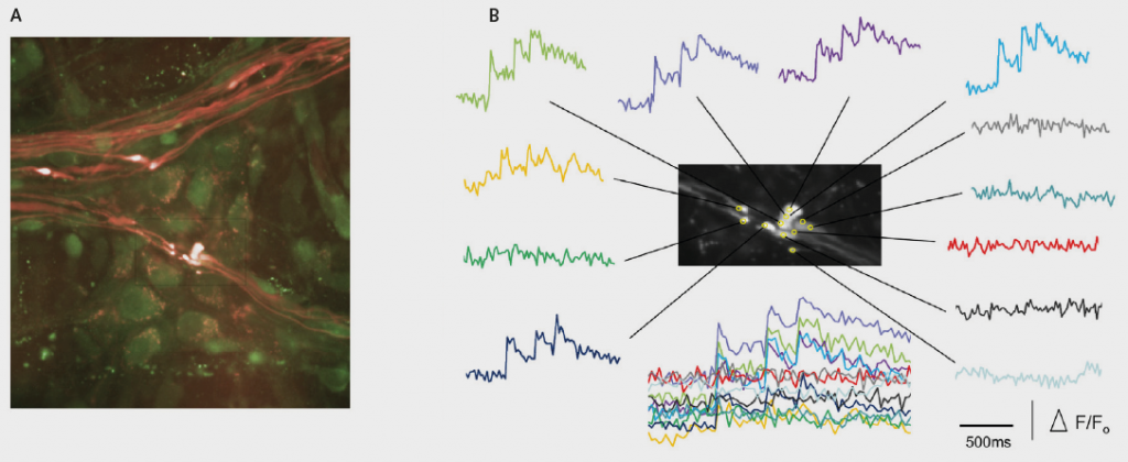
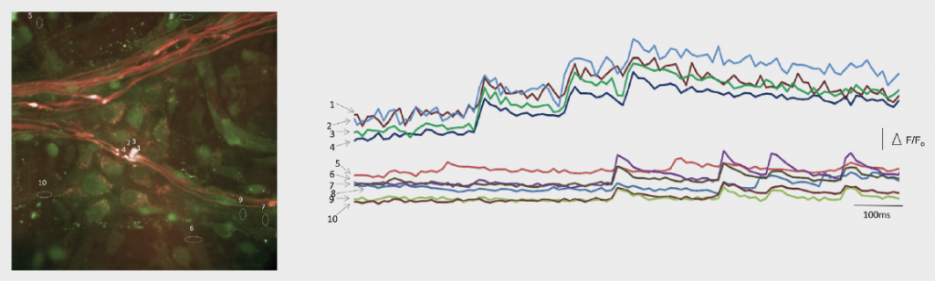
The team’s challenges were overcome with the Evolve® 512 Delta (new series now available). Using the camera for calcium imaging enabled them to record from multiple sites simultaneously along single axons of interest. They were also able to ensure that nerve endings were recorded accurately by stimulating dorsal nerve roots and demonstrating that those calcium transients were evoked in the same nerve endings that were activated by stretch or acid. This work has recently been published.
Of significance, due to the sensitivity of the Photometrics range of EMCCD cameras, the team has been able to perform the first and only recordings from nerve endings that detect pain from internal organs.
They are now answering many new and exciting questions using this technique. “We have been stunned by the incredible sensitivity of the EMCCD cameras produced by Photometrics,” states Dr. Spencer. Also, the high frame rate acquisition of the Evolve Delta camera at full resolution is just what was needed to record dynamic physiological activation of fine nerve endings.