
Dr. Alexandre Fürstenberg
School of Chemistry, University of Geneva
Background
The lab of Dr. Alexandre Fürstenberg deals in the development and application of optical spectroscopic and microscopic tools, with a focus on single-molecule imaging using smart fluorescence probes.
Dr. Fürstenberg told us of his lab’s recent research, “We are interested in developing fluorescent probes… we use probes that are sensitive to the lateral pressure in biological membranes, they can tell you about the composition of the membrane, the packing of the lipids, these are mechanosensitive fluorophores that are called flippers… there is a new version of these flippers that blink, and that’s exactly what we need for super-resolution microscopy.”
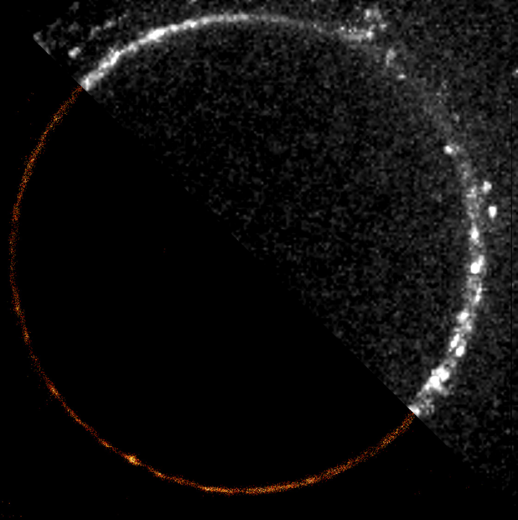
“Using these fluorescent flippers we want to look at the membranes of giant unilamellar vesicles (GUVs) and demonstrate super-resolution imaging with dSTORM.”
Challenge
Imaging GUV membranes at super resolutions levels is a demanding application for a scientific camera, due to the individual GUV cross-sections (with membranes filled with flipper probes) having low signal, and requiring many images over time to build up a dSTORM image. In addition, a large FOV is needed to contain larger GUVs so that tiling and stitching can be avoided, and high sensitivities are needed to detect fluorophores within the membrane.
Dr Fürstenberg discussed a previous camera solution, “I had an EMCCD camera already but it was 10 years old and too slow… we were really limited by the field of view, we wouldn’t have been able to look at the really large GUVs… we also need the sensitivity to look at individual probes at the GUV membranes which have very weak signals and which we could not really resolve before in certain lipid environments.”
I am very impressed by the different modes of the Kinetix, and its ability to go well beyond 1000 fps.
Solution
The Kinetix CMOS meets all the needs for this application, with a large FOV, high sensitivity thanks to 95% peak QE and minimal read noise, and high-resolution thanks to a large 10 megapixel sensor with a balanced 6.5 μm pixel size.
Dr. Fürstenberg outlined his experience with the Kinetix, “I needed a new camera so I went for the best one, I can see many advantages such as the really large field of view. We can do a factor of 10 better in terms of speed than the previous camera, this will help us for planning future experiments.
“It was pretty easy to set up the Kinetix thanks to the help from Photometrics staff and the camera also runs very smoothly with MicroManager, this was all a big selling point… It’s also great not to worry about having to center the region of interest as well, before we had to position our samples exactly at the center of the field, this problem is gone now with the Kinetix.”

Reference
José García-Calvo, Jimmy Maillard, Ina Fureraj, Karolina Strakova, Adai Colom, Vincent Mercier, Aurelien Roux, Eric Vauthey, Naomi Sakai, Alexandre Fürstenberg, and Stefan Matile, (2020), Fluorescent Membrane Tension Probes for Super-Resolution Microscopy: Combining Mechanosensitive Cascade Switching with Dynamic-Covalent Ketone Chemistry, Journal of the American Chemical Society 2020 142 (28), 12034-12038, DOI: 10.1021/jacs.0c04942