Prof. Xue Han and Dr. Eric Lowet
Biomedical Engineering Department, College of Engineering, Boston University
Background
The lab of Prof. Xue Han uses optical methods to probe neural circuits in awake animals. With a background in optogenetics, Prof. Han has developed many key molecular tools for optogenetics, including SomArchon, an archaerhodopsin-based voltage sensor. With a combination of voltage imaging using SomArchon, calcium imaging, and optogenetics, the Han Lab can optically interface with neural circuits.
Prof. Han told us about her experience with voltage imaging, “By using genetically encoded voltage sensors (GEVIs) we can record from many neurons in vivo, with their genetic identity and morphology. We can also observe sub-threshold dynamics separate from the firing of action potentials, we can probe the dynamics of individual neurons or a whole neuronal population. We can really see the voltage inputs now, which we haven’t been able to see over the past century.”
Through voltage imaging, Prof. Xue Han and postdoc Dr. Eric Lowet can investigate functional behavior from neural samples.

Challenge
The challenge of voltage imaging is speed, as described by Prof. Han, “How can you image a large area at high speeds? For voltage imaging we want kilohertz, 10s of kilohertz. A single action potential has a duration of one or two milliseconds, we are not going to be happy to record at one kilohertz, that gives us one data point for a millisecond event, and we may even miss it. The key challenge here is to image a large field of view at super high speed, as fast as the camera can go, as high as the computer can handle.”
At such high imaging speeds very low exposure times are necessary, resulting in low signal levels. As well as having a large field of view and extreme imaging speeds, the camera is also required to be highly sensitive with low noise levels, in order to record as much relevant data as possible over short time-frames of activity in neural samples.
We can see high signal-to-noise voltage from hundreds of neurons at once, the Kinetix is the first camera that was able to do this. This will definitely be a game changer in the voltage imaging field.
Solution
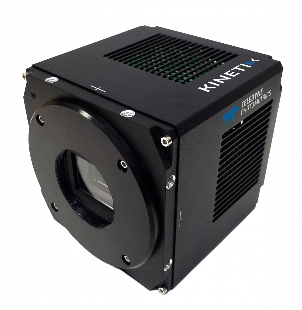
The Kinetix features the extreme speeds, large-format sensor, and high sensitivity required for voltage imaging, and removes the camera as a bottleneck for the imaging system. With the 8-bit Speed mode, the Kinetix can record at 500 Hz across all 10 megapixels, with far higher speeds at smaller regions, easily into the kilohertz and beyond.
Dr. Eric Lowet described a recent experiment with the Kinetix for voltage imaging, “We are imaging the visual cortex of an active, awake mouse at 40x, using the full field of the Kinetix. We can focus our laser point on a single neuron and see high signal-to-noise voltage spikes, which showed us that this 8-bit Speed mode does work, and I think this is awesome.”
“The most surprising thing for us was that we are able to record full field at 500 Hz and we were able to see single spikes and good signal to noise membrane voltage of neurons, which is very promising. Compared to other cameras we tested [the Kinetix] is the first camera that was able to do this, and with high quality.”
“We are able to record many hundreds of neurons at once or record few neurons at very, very high sampling rates. This opens up a lot of new opportunities, recording from many neurons will definitely be a game-changer in the field.”

