Dr. Jonathan Rocheleau
Percy Edward Hart Professor in Biomedical Engineering & Associate Director, Research, University of Toronto
Background
Jonathan Rocheleau is an Associate Professor in the Institute of Biomaterials & Biomedical Engineering at the University of Toronto. His laboratory uses high content imaging to study pancreatic islet biology.
Dr. Rocheleau told us, “We do high content microscopy because we’re interested in the idea that cellular metabolism is heterogeneous – that some beta-cells are metabolically poised for proliferation or survival, while others are poised to secrete insulin.” Dr. Rocheleau continued, “We’ve created a high content microscope for measuring anisotropy sensors that we refer to as Apollo. We started with a sensor that responds to NADP+ but believe that there’s a lot of potential for the development of many more sensors using this design.
Challenge
The use of a camera with very stable readout and noise characteristics is critical to precise measurements in this application. Dr. Rocheleau explains, “Anisotropy imaging is very precise. It’s a photophysical measurement we’re making. With other sensors you often see data shown as representative traces. The precision of anisotropy imaging allows us to collect and collate data across many different cells with little to no drift between experiments”
Smaller fields of view on previously used cameras limited the number of events that could be recorded in a single experiment. “When we go to 63x with cameras with smaller chips, we were only imaging tens of cells at a time. We need to do populations of thousands of cells, and you’d have to tile a lot of images together. When you’re tiling and having to image, all of that takes time.”
We like that the Iris 15 has such a big chip. The camera gives us so much information, allowing us to collect tons of data.
Solution
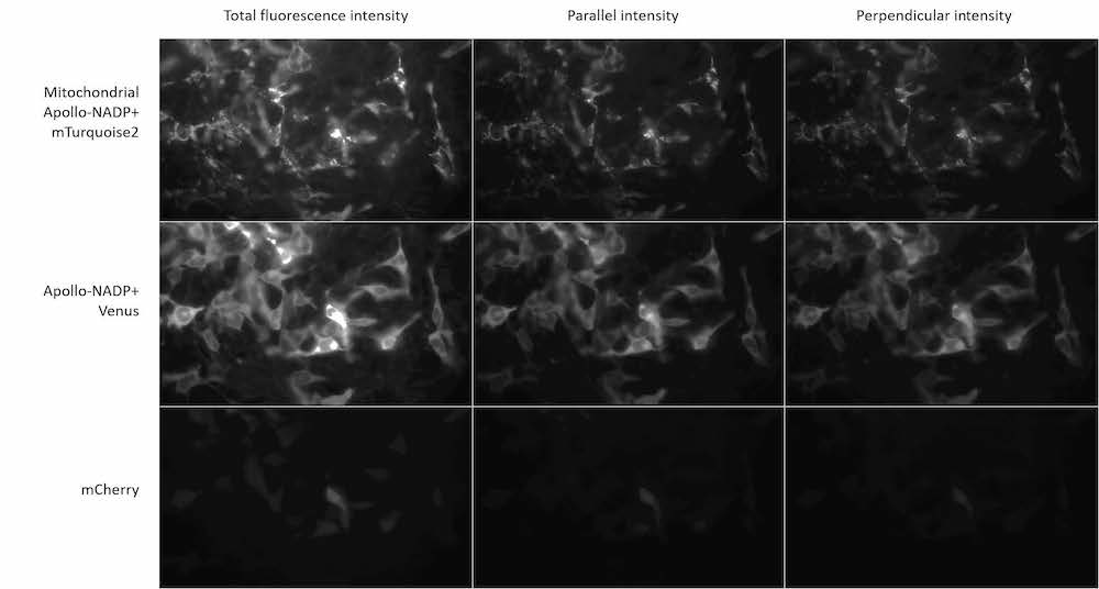
The 25 mm field of view of the Iris 15 makes it a great camera for high content imaging applications, allowing Dr. Rocheleau’s team to image more cells and remove the need for tiling when projecting multiple images onto a single sensor. Dr. Rocheleau told us, “We like that the Iris 15 has such a big chip. In the future, we want to collect both parallel and perpendicular fluorescence simultaneously on the same chip. The camera gives us so much information, allowing us to collect tons of data.”
Additionally, the Iris 15’s noise stability and sensitivity across a wide spectrum of wavelengths allows the lab to use fluorescent sensors of a variety of emission wavelengths to tailor their imaging needs. Dr. Rocheleau concluded, “With this sensor we get very precise measurements. The precision of measurements is very high day to day, and we can make comparisons between experiments very well.”