Dr. Jan Huisken, Director of Medical Engineering
Dr. Kurt Weiss, Postdoctoral Researcher
Morgridge Institute for Research, Wisconsin
Background
Dr. Jan Huisken, Director of Medical Engineering at the Morgridge Institute for Research has been instrumental in the invention and development of Light Sheet Microscopy. One of his current research interests is in the development of novel methods to image cleared tissues.
Dr. Kurt Weiss, postdoctoral researcher in the Huisken lab, told us, “We image entire organisms and organs that have been optically cleared. That includes samples from fish to mouse organs in the range of 1-20 mm. As a microscopy development lab with no eyepieces on our microscopes we depend exclusively on scientific CMOS cameras for feedback,” Dr. Weiss continued, “One of the common features of our samples is that they are fairly large, so we look for low magnification, and a moderate numerical aperture in our setup.”

Imaged in 2 tiles using the full field of view of the Iris 15 Scientific CMOS camera. 1 µm per pixel resolution. Color coded for depth.
Challenge
For their light sheet imaging experiments, large fields of view are highly desired to minimize the number of images required to image a whole specimen. Dr. Weiss explained, “Most objectives have at least a 21 mm field number so the limiting factor is generally the camera.” Additionally, processing images can become challenging when smaller sensor sizes limit the field of view in a tiled image. “Smaller chips and larger pixels forced us to use a large number of image tiles, increasing imaging time and post-processing complexity.” Dr. Huisken adds, “At the same time we want to achieve diffraction limited resolution across the entire field of view. Hence, a large sensor with small pixels is ideal for light sheet imaging of cleared specimens.”
The Iris 15 camera has both a large chip and a small pixel size so we can optimize the numerical aperture of our lenses. The 4.25 µm pixel size on the Iris 15 allows us to use more of the 0.28NA at 4× than other cameras without zooming.
Solution
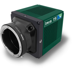
The large, 25 mm sensor coupled with small, 4.25 µm pixels make the Photometrics Iris 15 scientific CMOS camera a perfect fit for the light sheet imaging of cleared tissues at the Huisken lab.
Dr. Weiss told us, “The Iris 15 camera has both a large chip and a small pixel size so we can optimize the numerical aperture of our lenses. The 4.25 µm pixel size on the Iris 15 allows us to use more of the 0.28NA at 4× than other cameras without zooming. I was surprised with the sensitivity. I know that physically a small pixel will gather fewer photons per unit area. Thus, I expected to take a significant hit in sensitivity, but we can simply increase laser power slightly and get just as good of a signal as larger pixel cameras.” Additionally, the speed of the Iris 15 allows for extremely fast light sheet imaging experiments. Dr. Weiss shared, “We use this camera on a custom light sheet microscope to obtain 1-2 micron resolution across 10 mm samples in a matter of seconds.”

