
Greg Perry
Image Resource Facility, St Georges, University of London
Dr. Ferran Valderrama
Academic Director of the Image Resource Facility
Dr. Dan Osborn
Molecular and Clinical Sciences Research Institution
Background
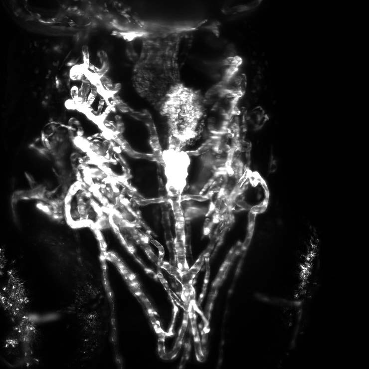
Greg Perry, from the Image Resource Facility at the University of London, works closely alongside academics such as Dr. Osborn and Dr. Valderrama, to improve imaging across a variety of research applications including long-term live imaging of zebrafish development and 3D organization of prostate cancer cell structures.
The Osborn lab is interested in translational research using zebrafish embryos as a model organism for tissue regeneration. The group manipulates an enzyme called nitrogen reductase that can be used to convert a pro-drug into a cytotoxic agent, permitting temporal control of apoptosis in these model organisms. Once apoptosis has been initiated, the regeneration of tissue can be observed using microscopy to elucidate mechanisms of repair.
Dr Valderrama’s group are interested in improving the understanding of prostate cancer progression. Prostate cancer originates from lesions in the glandular structures (acini) of the prostate. The group has developed a 3D cellular model that recapitulates the morphogenesis of these acini and serves as a model for cancer formation. The group uses this model in conjunction with microscopy methods to understand the molecular mechanisms of prostate cancer progression
Challenge
Cleared samples are typically very large which means that low magnification is often necessary to image the entire sample. However, this comes at the cost of resolution.
The Image Resource Facility recently acquired a new OpenSPIM (T-SPIM) system, which will be used to further advance the research areas of the Osborn Group and Valderrama Group. The new OpenSPIM setup permits live imaging of whole organisms such as transgenic zebrafish to enable the visualization of tissue repair over the course of days, as well as allowing the imaging of 3D acini cellular structures.
The facility required a camera to use alongside this new system to maximize the information gained during acquisitions. The OpenSPIM system requires a camera that can offer high speed, sensitivity and a large FOV to facilitate fast image capture of these large 3D samples. Sensitivity is important not only for imaging, but also for alignment which can be tricky with dim samples.
The camera is ideal for our OpenSPIM system due to the sensitivity and the large field of view
Solution
Greg Perry is now using the Teledyne Photometrics Iris 9 on their new OpenSPIM system to collect time-lapse images of 3D model systems alongside collaborating groups such as the Osborn Group and Valderrama Group.
Mr Perry told us that, “[The Teledyne Photometrics Iris 9] is a really good camera that is clearly very flexible in application”. He also shared that, “The camera is ideal for our OpenSPIM system due to the sensitivity and the large field of view.” Mr Perry also noted that the camera was very fast which is ideal for their applications.