
Dr. Rui Ma, Prof. Jochen Guck
Max Planck Institute for the Science of Light (MPL), Germany
Background
Dr. Rui Ma is working on the development of optical microscopy systems at MPL, and is currently involved
with building a light sheet microscope in order to image zebrafish, a biological model system used by
colleagues at MPL to study spinal cord injury.
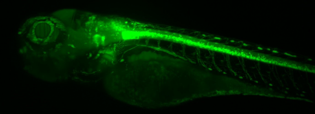
This imaging is done with genetically modified zebrafish that express a fluorescent protein, such as GFP.
These fish are imaged with fluorescent light-sheet microscopy at 10x in order to image the entire fish, but
Dr. Ma has plans to image at higher magnifications with higher NA in order to image the spinal cord in more
detail.
Using a custom-built OpenSPIM-based light-sheet microscope, image stacks of zebrafish are taken at
different angles at different time points, and the samples are monitored for several days.
Challenge
Live zebrafish make for large and dynamic microscopy samples, meaning an imaging system would need a sufficient field of view to capture an entire sample.
As well as a large field of view that matches up with a light sheet system that works with large samples, sensitivity is needed in order to image a range of different fluorophores at different wavelengths, requiring a camera with a high peak QE as well as a broad sensitivity across the visible spectrum.
This light-sheet system was built with the Kinetix in mind, we wanted to use the larger field of view, higher QE and faster framerates, compared with other cameras.
Solution
The Kinetix sCMOS is the ultimate solution for light-sheet microscopy, featuring a huge 29.4 mm field of view, combined with high speeds, high resolution, and sensitivity from 200-1000 nm range.
Dr. Ma uses the Kinetix with her custom-built light sheet system, and described her experience, “This system was built with the Kinetix in mind, we wanted to use the larger field of view, higher QE and faster framerate, compared with other cameras.”
“The Kinetix matches well to our light-sheet application, the large field of view is a big benefit for us, especially if we want to image our large samples without stitching.”
“Overall the Kinetix is a good camera. The zebrafish is in the center of our field of view and we can crop the sensor to capture it, this reduces the data size. We also tried other fluorescent proteins and the sensitivity is good for these too.”
