Prof. Suliana Manley, Dr. Luc Reymond, Dr. Willi Stepp, Mr Chen Zhang
Laboratory of Experimental Biophysics, EPFL, Lausanne, Switzerland
Background
The Laboratory of Experimental Biophysics (LEB) at EPFL, headed by Prof. Suliana Manley, develops and uses fluorescence imaging techniques combined with live-cell imaging and single-molecule tracking to determine how the dynamics of protein assembly are coordinated.
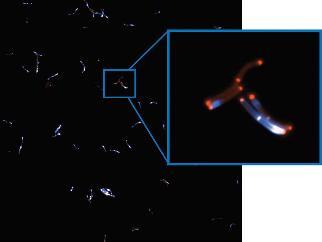
Dr. Stepp and Mr. Zhang are researchers in the LEB, with Mr. Zhang studying the cell cycle dynamics of bacteria using super-resolution imaging techniques such as STORM and SIM, while Dr. Stepp operates as a microscope and optics specialist, enhancing imaging systems and instrumentation. Dr. Stepp spoke about the LEB: “We try to get the right microscope technique for the problem at hand… people come to us with imaging problems and we try to solve it with any technique that is out there.”

Challenge
The LEB uses a range of different imaging systems and techniques, including custom-built microscopes providing specialized capabilities. Dr. Stepp outlined some of the detector needs at the LEB, “for our current project we can have issues expressing enough fluorophores in the bacteria, so there we need high sensitivity, especially as the experiments are performed on our iSIM, which is quite a photon hungry setup… for other projects on mitochondrial dynamics, we also need speed and field of view… if we have one camera that could do it all that would be the best.”
The camera needs for each imaging system and each type of sample are varied in terms of resolution, sensitivity, field of view, and speed, meaning the ideal solution would be a highly flexible detector that can move between different imaging systems, yet is powerful enough to deliver sufficient acquisitions across these different systems.
We had [the Kinetix] set up on our system pretty quickly and our first images were great, very impressive.
Solution
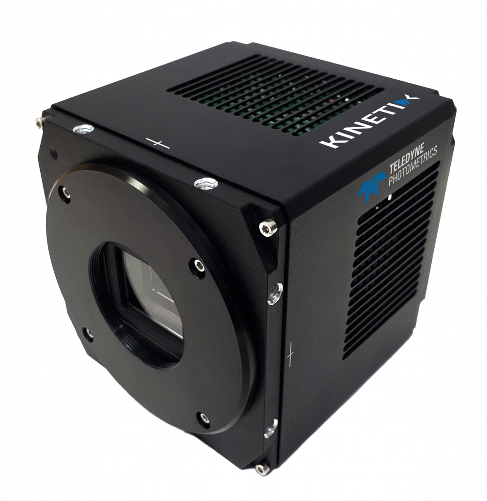
The Kinetix CMOS is a highly powerful camera that can be easily adapted to a number of different imaging systems, featuring a large 29 mm field of view with a balanced 6.5 um pixel size, a very high speed of over 500 fps full frame, all with low read noise and high quantum efficiency.
Dr. Stepp discussed his experience with the Kinetix, “We have a number of imaging systems that would be suitable for the Kinetix… the speed was interesting paired with the big field of view as our samples have short time ranges.”
“The build quality is great, the mounting feels solid, and the initialization is faster than our other cameras so that makes imaging faster on startup in the morning… We had [the Kinetix] set up on our system pretty quickly and our first images were great, very impressive.”

