
Prof. Francesco Pasqualini
Synthetic Physiology Lab, University of Pavia, Italy
Background
Prof. Francesco Pasqualini is a Harvard-trained bioengineer leading the synthetic physiology laboratory at the University of Pavia, currently researching cardiac development using engineered cell culture platforms. By optimizing this platform with cell lines and then moving to human induced pluripotent stem cells, Prof. Pasqualini can get a unique perspective of the developing heart.
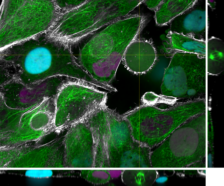
Prof. Pasqualini told us more, “We are studying the mechanobiology of the heart as well as investigating the use of various extracellular matrix components to control tissue and organ-level behavior. We are particularly interested in recapitulating the early phases of human heart development using defined extracellular matrix and cardiac cell types. All using live-cell microscopy, of course.”
This study uses a Nikon Ti2 microscope and a modular spinning-disk confocal module, the X Light V3 from CrestOptics.
Challenge
Cardiac research involves imaging of both structural and functional aspects of cardiac cells. While structural imaging requires a camera with a large field of view and a small pixel to get high spatial resolution at the desired magnification, functional imaging requires a camera sensitive enough to operate at high speeds to capture calcium and voltage activity across cells and tissues.
When working with multiple cell types and multiple timescales, it is vital to have a flexible yet powerful imaging system that can meet the needs of each experiment, whether imaging the morphology of large groups of cells or imaging small sub-populations with high speeds to observe activity.
The Kinetix does everything we had hoped for, and is flexible enough for our future experiments!
Solution
The Kinetix is a proven solution when paired with the Nikon Ti2 and CrestOptics X-Light V3, resulting in a powerful imaging system. This allows for flexible imaging both now, and in the future when bigger, faster, more sensitive experiments are planned to image ultra-low voltage signal levels in developing heart tissues in vitro.
Prof. Pasqualini described his experience with the Kinetix, “I like the different Kinetix modes, we can image lots of cells at ~100 fps, and if we need to, we can look at another type of cell at ~1000 fps for calcium imaging. I also like Sub-Electron mode for images that don’t need to be fast, the lower read noise makes a big difference.”
