
Prof. Jan Pielage
Department of Biology, RPTU University of Kaiserslautern-Landau, Kaiserslautern, Germany
Background
Professor Jan Pielage’s research focuses on identifying molecular mechanisms controlling synapse formation, function, and stability. Prof. Pielage told us more, “We use Drosophila models to identify novel genes that are required to maintain the stability or function of the neuromuscular junction (NMJ), the connection between nerve and muscle. We have identified a number of genes, and these are conserved and can lead to disabilities in mouse or human models.”
“We want to look at neurodegeneration across a whole Drosophila NMJ and see how the signal pattern changes, to learn more about potential compensatory mechanisms and disease progression.”
Challenge
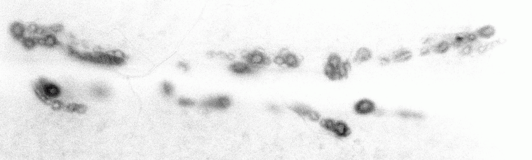
Prof. Pielage described some of the challenges this application involves, “We want to monitor potential defects in synaptic transmission at the resolution of single active zones in the NMJ, for this, we require extreme sensitivity as the signal is very low.”
“We also need high-speed imaging to identify these events in real-time, this is in the range of 10-100 Hz, but we also want to image voltage-sensitive dyes, and for this, we would need to go up to 1000 Hz. So, we need to really push the potential of the camera.”
“We also want to image the entire NMJ, it is critical to understand which release sites are active. Previously we relied on electrophysiology and could detect release events, but didn’t know where they occur. For this, we need optical methods.”
This application needs a combination of high speed and high sensitivity across a large field of view, in order to observe all the events across an entire Drosophila NMJ at sub-cellular resolution.
This system was designed for the Kinetix22, when we compared it to competitors the Kinetix22 really was the best for our application, it’s the perfect camera!
Solution
The Kinetix22 is the ideal solution for this application, and Prof. Pielage explained his experience with the Kinetix22, “This system was designed for the Kinetix22, the improvement in temporal resolution and sensitivity is critical. We were previously just at the detection limit but now it looks nice, we were surprised that we can see our signal. We can also look at the kinetics of these events, this is really a great tool and we compared it to competitors, the Kinetix really was the best for our application, it’s the perfect camera.”
“These experiments weren’t possible beforehand, now we can see miniature release events within the NMJ, something that we cannot achieve using electrophysiology. These optical recordings dramatically improve the dimensions of our research, and if we then correlated directly with electrophysiology and immunochemistry, we can really gain novel insights into these processes.”
“It works well in MicroManager, and the setup was very easy, it works from the get-go.”
