
Dr. Rory Power
Advanced Light Microscopy, EMBL Heidelberg, Germany
Background
Dr. Rory Power is a staff scientist and engineer at the advanced imaging center of the EMBL headquarters in Heidelberg. Involved in a variety of projects, Dr. Power also oversees Ph.D. students involved in building custom light sheet imaging systems.
Dr. Power described their light sheet imaging system, “It’s an oblique plane microscope (OPM) that uses a single objective, which allows us to do light sheet microscopy in a more traditional inverted, epifluorescence setup with less restrictions on sample geometry and using a water dipping objective.”
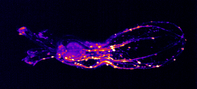
“This system is for imaging of a little tentacle monster Nematostella, which are interesting from a morphological and behavioral point of view, how their muscle hydraulics and neural dynamics influence their development and motion. We are using our light sheet system to image contractile waves of motion within these animals.”

Challenge
Dr. Power told us about the imaging challenges he faces in his work, “The Nematostella cannot be constrained, these are fully grown animals that need to move around and undergo normal behaviors. These samples are moving freely in a droplet, so we only image them when they move into the camera’s field of view. We also want to go fast so we can capture dynamic motion while retaining a large FOV.”
In addition to this, Nematostella is a light-sensitive organism, meaning a low light regime is needed, reducing signal levels and requiring a highly sensitive camera. Due to their size (~1.5 mm in length, ~200 μm in width), the Nematostella can only be imaged with a light sheet when the body axis is in a certain orientation, further requiring a large FOV to increase the number of good imaging events.
Finally, in order to further increase the FOV, a low magnification objective is used which inherently has a lower numerical aperture, which can be challenging when using an OPM imaging regime.
The high speed and the large chip of the Kinetix made it ideal for our imaging applications.
Solution
The Kinetix is an ideal solution for this imaging application, featuring both a very large 29 mm FOV combined with a very high speed of 500 fps across this entire sensor. The Kinetix is widely used for light sheet and means researchers no longer have to compromise.
Dr. Power described his experience with the Kinetix “The speed and the large chip of the Kinetix were the things that made it ideal for our application. Right now, the full speed of the Kinetix hasn’t even been leveraged in recent experiments, we can easily go a factor of 5x faster.”
“Software setup was absolutely fine for capturing images when triggered, everything worked so no problems. Introducing the camera to the system hardware was easy.”
The Kinetix sCMOS replaced a traditional sCMOS camera used for alignment/testing due to the better performance and allows for high-resolution imaging across a large FOV while maintaining a high speed.
Reference
Singh R., Subramanian K., Power R.M., Paix A., Ikmi A., Prevedel R. (2022) An oblique plane microscope for mesoscopic imaging of freely moving organisms with cellular resolution, bioRxiv 2022.07.15.500249; doi: https://doi.org/10.1101/2022.07.15.500249
