Dr. Christian Simon, Mr. Florian Gerstner
Carl-Ludwig-Institute for Physiology, Leipzig University, Germany
Background
Dr. Christian Simon and PhD student Mr. Florian Gerstner are involved in neuroscience research, in particular investigating spinal cord sections and motor neuron functionality in mouse models.
Dr. Simon described his work, “For the spinal cord, if you break it into small sections, you cut off all the dendritic trees and this doesn’t really work, we are using a new technique where you divide the spinal cord into very thick sections, where you can visually target the surface motor neurons and still have the circuitry intact.”
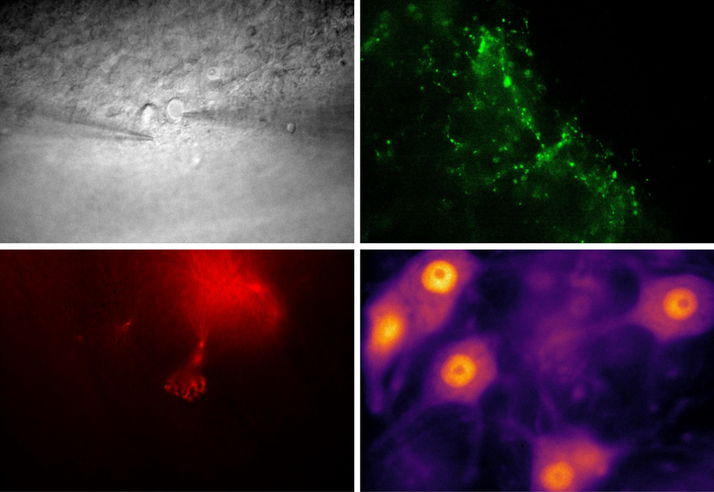
Mr. Gerstner told us more, “We are doing a lot of electrophysiology on these thick samples, primarily patch clamp recordings, but we are also doing fluorescence and DIC microscopy to target certain cells and check their viability. We are using GFP and Atto dyes towards red.”
Challenge
For thicker samples a high sensitivity camera is vital due to the scattering that occurs for both DIC and fluorescence, as well as high sensitivity across a range of wavelengths, considering the use of traditional visible GFP and another dye further towards infrared that better suits thick samples.
Dr. Simon had a previous camera that lacked sensitivity, “With our old camera the sensitivity wasn’t sufficient, especially in these thicker tissues and in DIC. We are targeting cells beneath the surface, so we need good image quality, we have to image the layer beneath to get more intact cells.”
Mr. Gerstner outlined more challenges, “We are using two types of objective, a 10x to locate the area we want to see, then for patching we use a 60x in order to target individual cells. Our previous camera imaged at around 30 fps which was low for us, we need more speed in order to get the patch pipette to the desired cell.”
Overall, this application requires high and broad sensitivity, as well as high speed and a pixel size suited to high-resolution imaging at a range of different magnifications.
The Moment is an easy-to-use plug and play camera that produces quality images with a good time resolution. We didn’t have any problems.
Solution
The Moment CMOS is a high-speed, easy-to-use camera with high sensitivity in both the visible and near-infrared regions. With a small 4.5 µm pixel, the Moment is well suited to both low-magnification localization and high-magnification sub-cellular imaging.
Mr. Gerstner described his experience with the Moment, “It’s easy to use, since with Micro-Manager its plug and play with the single cable, it’s a neat way to do things. It’s been a comfortable camera to use, that produces quality images with a good time resolution. It works perfectly with great resolution at both of our magnifications. We didn’t have any problems.”
Dr. Simon told us more, “[The Moment] improved our approach since we can do everything within one software. With our previous camera fluorescence was horrible, but now even with thick samples we are all very happy.”