Friederike Seifert, Dr. David Fleck, Prof. Marc Spehr
Department of Chemosensation, Insitute for Biology, RWTH Aachen University, Germany
Background
The lab of Professor Marc Spehr is working on the olfactory system of mammals and the underlying mechanisms of pheromone detection. Pheromone detection is key for social communication in the animal world.
The main methods of choice utilized by Dr. David Fleck and Ph.D. student Friederike Seifert are electrophysiological techniques, in combination with imaging to visually observe the signal transmission in the vomeronasal organ (VNO) upon very sophisticated stimulation with pheromones. The imaging techniques range from simple backfilling of neurons, identifying subsets of neurons expressing certain types of receptors, to measuring activity by detecting signals from genetically encoded (GECI) or dye-based Ca2+ indicators.
Challenge
For a typical experiment, a slice of the VNO is mounted under a microscope with the aim to record an individual neuron using electrophysiology, with a patch pipette. Typically, near-infrared (NIR) illumination is utilized to focus on the tissue and the pipette in order to achieve high contrast. For this purpose, special coatings had to be used on older CCD cameras in order to get a sufficiently high sensitivity – this compromised sensitivity in the visible spectrum relevant for the imaging of fluorescent reporters and dyes.
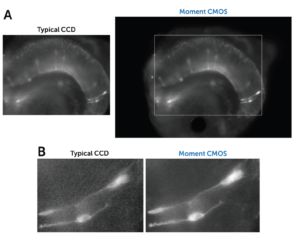
Additionally, existing detection solutions had very small sensor areas resulting in being able to just observe a small fraction of the entire VNO sample. This also made it very difficult to make conclusions about the overall context and anatomy of individual cells.
I´d like to buy one more, please!
Solution
The Moment CMOS is an ideal solution for this demanding application, overcoming the problems a classically used patch-clamp camera had faced. The general sensitivity of the Moment between 200 1100 nm allows the camera to have excellent sensitivity in both the visible range and the NIR, without compromise. Sensitivity is much improved compared to the previous CCD solution, resulting in better signal-to-noise ratios, whether for brightfield visualization of the patch pipette or fluorescence imaging of individual neurons.
The Moment´s higher maximum speed of 50 fps gives more instant feedback while positioning the pipette, but also enables a higher recording speed of fluorescent and/or dye-based dynamics opening new insights into physiologically relevant processes.
With the much larger diagonal field of view of 17 mm, the members of the Spehr Lab can finally detect the VNO in a single field of view, resulting in a much higher throughput and more efficient sample handling, allowing for more experiments. This results in a system that is both simpler and more powerful.
Lastly, the ultra-compact size of the Moment (40 x 42 x 50 mm) and the extremely low weight (96 g) allow for easy integration into electrophysiological imaging systems, and the global shutter triggering plus passive cooling makes it perfectly suited for upright microscopes typically used for electrophysiological experiments.