Naoki Mochizuki, Director
National Cerebral and Cardiovascular Center,
Department of Cell Biology
Background
The team at Department of Cell biology at the National Cerebral and Cardiovascular Center is focused on exploring the molecular mechanism by which blood vessel formation and cardiogenesis are regulated during development. To meet their research goals, the team must determine how cardiovascular development is precisely regulated.
Understanding of the development of cardiovascular system contributes to developing new strategy for cardiovascular regeneration. Currently the team is using zebrafish to investigate cardiovascular development. Zebrafish provide great advantages with quick development, extra-embryonic development, translucency and easy gene manipulation. The embryos expressing fluorescent proteins under the cardiovascular-specific promoters can be visualized as they are developing.

Challenge
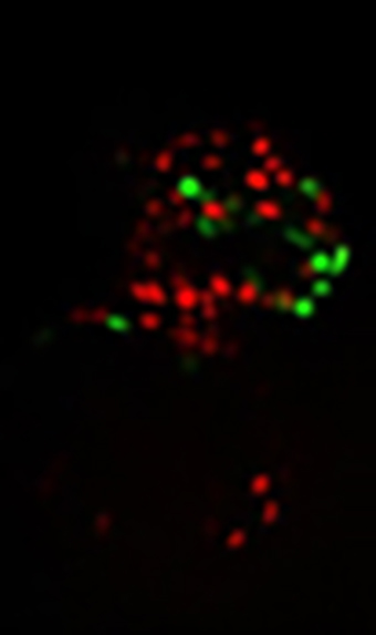
The only way to obtain the high speed needed (video rate image of fluorescence), is through the combination of CCD and spinning disk confocal microscopy. The imaging solution used must support the ability for high speed image acquisition of live zebrafish embryos expressing green fluorescence and red fluorescence.
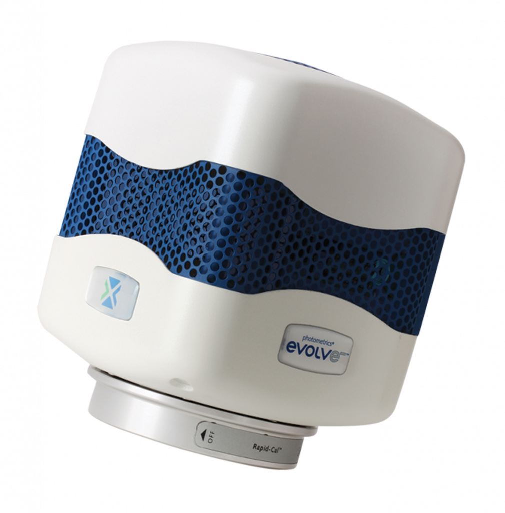
The quality and speed of the Evolve 512 EMCCD camera has enabled us to capture zebrafish embryo developments under challenging conditions.
Solution
Having used the CoolSNAP HQ2 CCD from Photometrics in the past, the decision was made to pursue a more advanced solution from the same company. The team selected the Evolve® 512 EMCCD camera (new series now available). “The quality and speed of the Evolve 512 EMCCD camera has enabled us to capture zebrafish embryo development under challenging conditions,” states Mr. Mochizuki.

Further Information
Additional information about the Department of Cell Biology at National Cerebral and Cardiovascular Center is available at: http://www.ncvc.go.jp/english/res/str_ana.html
