
Dr. Deepak Nair
Indian Institute of Science, Centre for Neuroscience
Background
Research at the Nanoorganization Lab, Centre for Neuroscience, Indian Institute of Science in Bangalore India borders on the interface of single molecule spectroscopy and molecular and cellular neuroscience. Here, team members develop and adapt state of the art paradigms in ultra-high and super resolution microscopy to image molecules at the synapses of living neurons.

Upper Panel: The epifluorescence image of a COS7 cell stained with tubulin antibody (A) which is then revealed by a secondary antibody coupled to the fluorophore Alexa 647. and Corresponding super resolution image (B).
Lower Panel: Gallery of epifluorescence images of different regions (C, E, G, I) from the cell and their corresponding super resolution images (D, F, H, J).
Challenge
The research team is interested in the molecular and cellular mechanisms that mediate basal synaptic transmission and plasticity. The routine imaging techniques used include FRAP, Photoactivation, Single Molecule Tracking and Stochastic Super Resolution imaging.
The team was interested in a widefield detection system with a high dynamic range that could handle both ensemble and single molecule fluorescence. Single molecule tracking in live cells is challenging due to heterogeneity in the signals and auto fluorescence from cells which make the contrast difficult to achieve. Most importantly the data needs to have good signal to noise ratio.
The Evolve is great for stochastic super resolution microscopy and meets our expectations in both ensembles and single molecule microscopy.
Solution
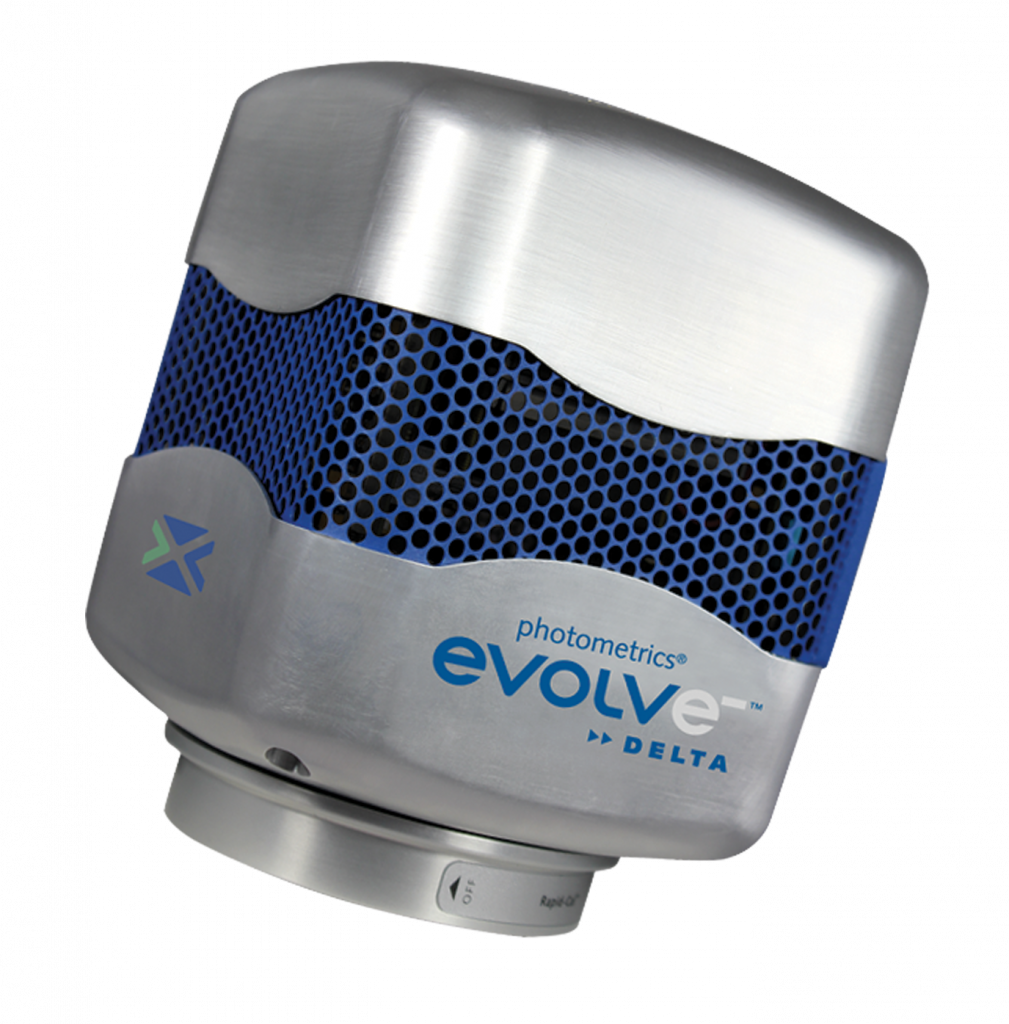
The Evolve® 512 Delta (new series now available) was recommended and having used a prior version of the Evolve camera, the team was happy to integrate a newer model into their system. They had also previously used TwinCam (Cairn Research) to implement a multichannel imaging system to acquire spatially similar but spectrally different images at the same time.
“We were interested in the Evolve Delta for its low read out noise, sensitivity and ability to image very high frame rates per second,” Dr. Nair shares. By coupling the dual camera detector unit with a total internal reflection microscope (Azimuthal TIRF, Roper France), they were able to meet expected demands.
“The Evolve cameras also gave us the ability to interface with third party software to drive the acquisition, which made it optimal for a wide range of applications,” Dr. Nair continues. The microscopy configuration also enabled the ability to collect routine images in the widefield and single molecule regime. The camera’s sensitivity and dynamic range extended the microscopic capabilities from micrometers to tens of nanometers.
“The Evolve 512 Delta is great for stochastic super resolution microscopy and meets our expectations in both ensemble and single molecule microscopy,” Dr. Nair concludes.
Further Information
Additional information about the research lab at the Centre for Neuroscience at the Indian Institute of Science in Bangalore is available at: http://www.cns.iisc.ernet.in/deepak/