Dr. Fabian Voigt, Postdoctoral Researcher
Prof. Fritjof Helmchen
Brain Research Institute, University of Zürich
Background
The mesoSPIM initiative (mesospim.org) is an open science project aimed at making light-sheet microscopes for imaging large cleared tissue samples more accessible to the imaging community. The acronym mesoSPIM stands for “mesoscale selective plane illumination microscopy” and the project was started in 2015 by Dr. Fabian Voigt and Prof. Fritjof Helmchen at the Brain Research Institute of the University of Zürich.
The idea was to create a highly modular and versatile light-sheet microscope capable of quickly scanning cleared organs and entire organisms. The mesoSPIM can image a mouse brain with an isotropic resolution of 6.5 µm within 8 minutes and is compatible with all clearing techniques. Instructions and software to set up and operate the instrument are freely available, and 10 mesoSPIM instruments have been set up around the globe at a wide variety of institutions such as the Wyss Center Geneva and the Sainsbury Wellcome Center in London, with several more instruments under construction.

Challenge
For the next generation mesoSPIM, Dr. Voigt and team set out to make the setup more compact and cost-efficient by turning it into a benchtop instrument. Unlike its bigger brother, the benchtop mesoSPIM does not need a dedicated optical table which drastically lowers the required budget. In shrinking the system down to a 45 x 60 x 50 cm box, Dr. Voigt had to find an alternative to the zoom macroscope utilized in the original mesoSPIM – with an overall length of 600 mm and a weight in excess of 15 kg, it would be too large and too heavy for a benchtop mesoSPIM.
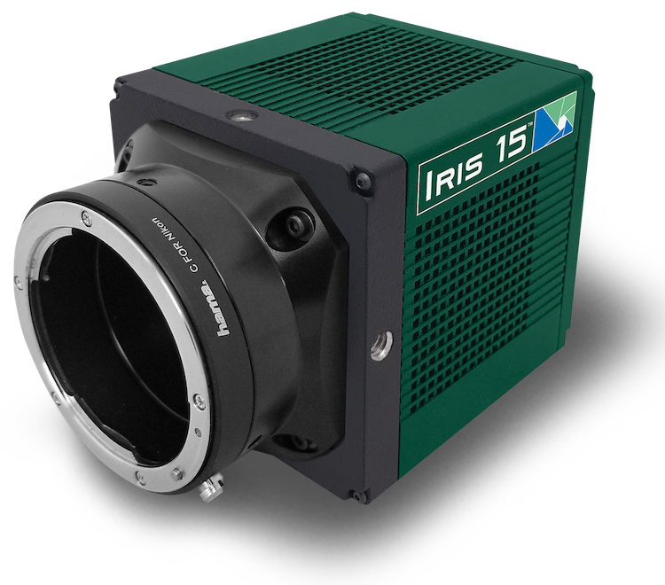
We tested the Iris 15 and found its large detector size and rectangular sensor ideal for our applications.
Solution
One option was to replace the combination of a zoom macroscope and a 4 MP sCMOS camera in the original mesoSPIM with a camera with a higher pixel count and a set of fixed-magnification objectives.
Therefore, we tested the Photometrics Iris 15 and found its large detector size and rectangular sensor ideal for our applications: For example, in combination with a 1.2x telecentric machine vision objective, the microscope has a FOV of 17.9 x 10.5 mm – perfect for imaging a whole mouse brain processed using the iDISCO clearing protocol. Owing to the large pixel count, fine features can be imaged without the need for time-consuming tiling acquisitions.
In addition, the Iris 15 offers a Programmable Scan Mode which is essential to achieving near-isotropic resolution in our datasets: By making use of a technique called Axially Scanned Light-sheet Microscopy (ASLM), the mesoSPIM is capable of <7 µm axial resolution across FOVs up to 20 mm. Lastly, the small form-factor of the Iris 15 greatly helps us to build a portable next-gen light-sheet microscope.

