
Dr. Steven Pittler
Director of the Vision Science Research Center,
University of Alabama at Birmingham
Background
Professor Steven Pittler, Director of the Vision Science Research Center at the University of Alabama at Birmingham, uses fluorescence imaging to study the diseases related to vision. Dr. Pittler explains, “Our Molecular and Cellular Analysis Core focuses on the investigation of a range of ocular diseases in the anterior and posterior segments of the eye. This includes common diseases like age-related macular degeneration and diabetic retinopathy, as well as less common diseases like retinitis pigmentosa. Our facility allows several investigators to image ocular tissues that are stained with different primary and secondary antibodies. Our goal is to develop a system that can cover the range of fluorescence wavelengths from 300 to 800 nm.”
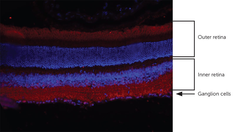
Photoreceptor cells (outer retina), bipolar, horizontal, and amacrine cells (inner retina) and ganglion cells layers. Image acquired with Micro-Manager 2.0 beta software on a Zeiss Axioplan 2, with DAPI (blue) and Cy5 (Red) filters at 20× magnification taken with the Iris 9 Scientific CMOS camera.
The primary antibody recognizes a channel protein in the photoreceptor outer segments and in the inner plexiform layer of the inner retina. Nuclear layers are stained with DAPI.
Challenge
Dr. Pittler and his colleagues must gather structural information from ocular tissues. For the Vision Science Center, this means distinguishing between various tissues and layers of the eye to learn about normal and disease processes in the eye. A high degree of spatial resolution is required. Additionally, sensitivity to detect low emitting areas of the sample is key to determining exactly which cells are expressing genetic abnormalities. Dr. Pittler told us, “The Iris 9 has outstanding sensitivity of detection and helped to minimize issues with autofluorescence.”
At 9MP resolution, the Iris 9 image quality is spectacular and allows for the capture of very fine detail
Solution
For the Pittler lab and the Vision Science Research Center molecular core, having the 4.25 µm pixel size and sensitivity over a wide range of visible wavelengths on the Iris 9 proved to be crucial to their retinal studies. “The resolution and level of sensitivity has made a huge difference. At 9MP resolution, the Iris 9 image quality is spectacular and allows for the capture of very fine detail,” Dr. Pittler continued, “Additionally, the fact that we can use this camera with Fiji and micro-manager was also an added plus.”