Prof. Naoki Yamawaki
Department of Biomedicine, Aarhus University, Denmark
Background
Prof. Naoki Yamawaki is a neuroscientist at Aarhus University, who told us about his research, “We are interested in understanding how stress affects the neuronal connections and communication in the brain, I am responsible for running the lab, doing experiments, mentoring, teaching students, and more.”
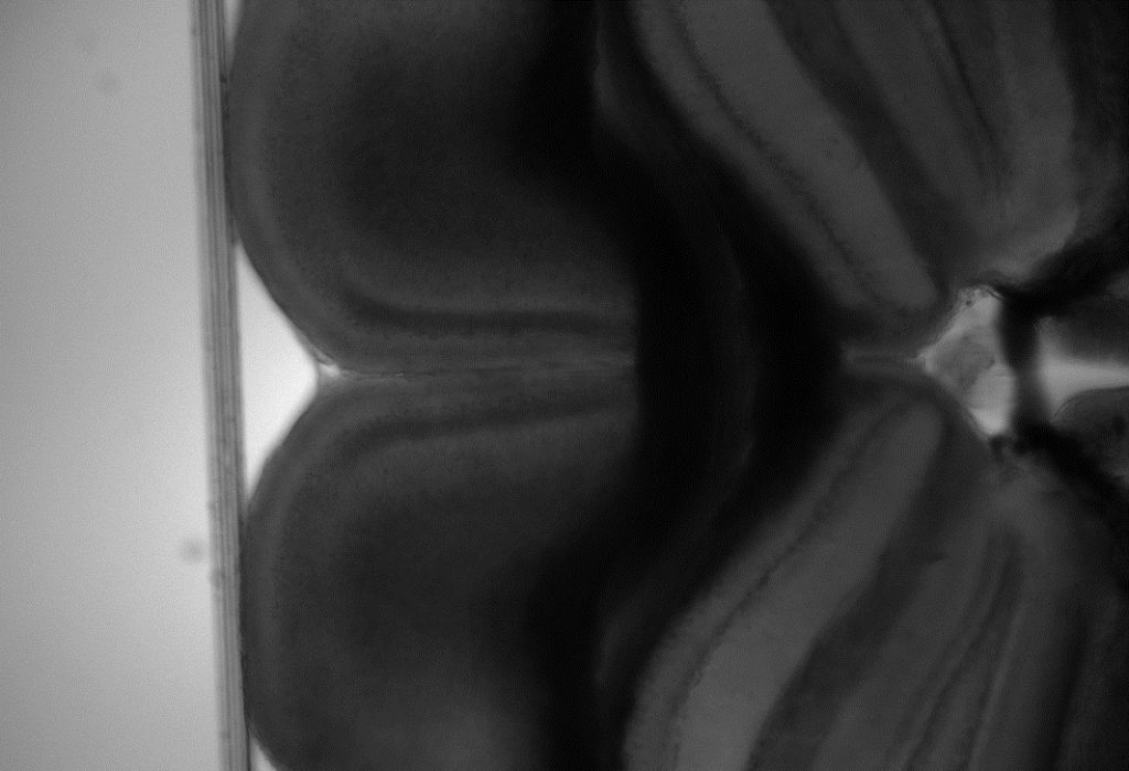
Prof. Yamawaki’s main research approaches include microscopy for imaging the brain and the neuronal circuits within, often using viral tracing and optogenetics combined with electrophysiology. These experiments are done on both healthy brains and brains in various disease states, in order to examine how certain neural networks act in these states.
Challenge
Patching tissue samples and using recording pipettes is a highly technical technique that brings challenges, both in order to patch a select area of the tissue, to ensure a good signal quality can be maintained during recording, and to avoid damaging brain tissue samples.
Prof. Yamawaki mentioned he had previously used a CCD camera; these devices often have limited speeds (less than 10 fps) and high levels of read noise (more than 10 e-), making it a challenge to image dynamic live samples or to detect low signals and image in low-light environments in order to avoid photobleaching.
A new imaging solution would need to feature both high speeds and high sensitivity, as well as high resolution and a large field of view in order to optimally image brain tissue slices and detect electrophysiological signals or fluorescently-labeled neurons. For this application, it was important to choose a flexible yet powerful camera.
The Moment has been working great, it is flexible, has low-noise, and was easy to install!
Solution
The Moment CMOS is the right tool when the goal is fast, high resolution imaging in a simple and highly compact package. The Moment achieves 50 fps across a 7 megapixel sensor, all with low read noise and a 17.5 mm diagonal field of view.
Prof. Yamawaki told us about his experience with the Moment, “We use the Moment for two things: ex vivo electrophysiology and fluorescence microscopy, in both cases we care about the sensitivity and resolution (bit depth and pixels) for imaging… In addition to sensitivity and resolution (chip size), I also considered the potential frame rate and controllability, especially its compatibility with NI data acquisition board (DAQ) and
open-source software.”
“The Moment has been working great so far, it is flexible, has low-noise, and was easy to install… [Teledyne] Photometrics staff were very helpful in every aspect such as determining the camera, purchasing, and troubleshooting.”