
Introduction
Electrophysiology is a method to study the function of electrically active properties of cells. It is widely used in both clinical applications, such as electroencephalograms (EEGs) and research applications, such as the monitoring of single ion channels. A large amount of the research on the physiology and electrical activity of neurons and their networks is performed at the cellular level, and knowledge of common microscopy techniques is essential.
The ability to visualize and capture images of the fine structures within tissues using an optical microscope has enabled researchers to uncover the secrets of how cells work and carry out further experimental techniques. Several of these techniques are incredibly useful for studying the function of neurons and related cell types such as in vitro patch clamp electrophysiology which aims to study ion channel activity of cells by making a secure seal between a glass micropipette and the cell membrane.
In neuroscience, advanced imaging methods like multiphoton microscopy have facilitated the visualization of structures deep into brain tissue and with very high temporal and spatial resolution. Further developments have also allowed for total optical interrogation of some cellular functions.
These applications require the user to have intimate knowledge and control of every component of the system. The selection of a camera for electrophysiology is of great importance to properly sample positioning and data collection. The Retiga ELECTRO from our QImaging CCD line offers electrophysiologists an ideal solution to imaging challenges commonly present with other cameras.
Imaging Challenges In Electrophysiology
Contrast Enhancements and Fluorescence
Many electrophysiology applications require the insertion of electrodes into cells or tissues but cells are often difficult to differentiate from the fine micropipette tip of the electrode. Differential Interference Contrast (DIC) microscopy, as well as other contrast methods like phase-contrast, are widely used to enhance the contrast between the edges of the sample and pipette. However, the pipette tip must be precisely placed within, or just on the surface of the sample to achieve proper data collection.The camera must, therefore, have high sensitivity and the ability to perform high contrast imaging.
The camera must also be capable of recording sample activity during stimulation. Many dyes used in electrophysiology are difficult to visualize because of low quantum efficiency at the fluorophores emission wavelength. Therefore, a camera with a high quantum efficiency over the entire visible wavelength range is also desired.
High-Speed Imaging
Cellular activity as a response to electrical stimulation or changes to membrane potentials can happen extremely quickly, especially in the field of calcium imaging. A camera with a frame rate capable of capturing these high-speed events must be carefully selected. Choosing a camera with a high maximum frame rate at full frame will ensure the capture of the highest number of data points while increasing throughput by maintaining a large field of view.
Noise, Vibration, and Interference
Electrophysiology applications often require minimal electronic noise and vibrations to acquire high-quality data. The electrophysiologist requires tight control of noise in the data so the selection of a camera is not only dependent on sensitivity and speed but also its ability to add as little noise to the system as possible.
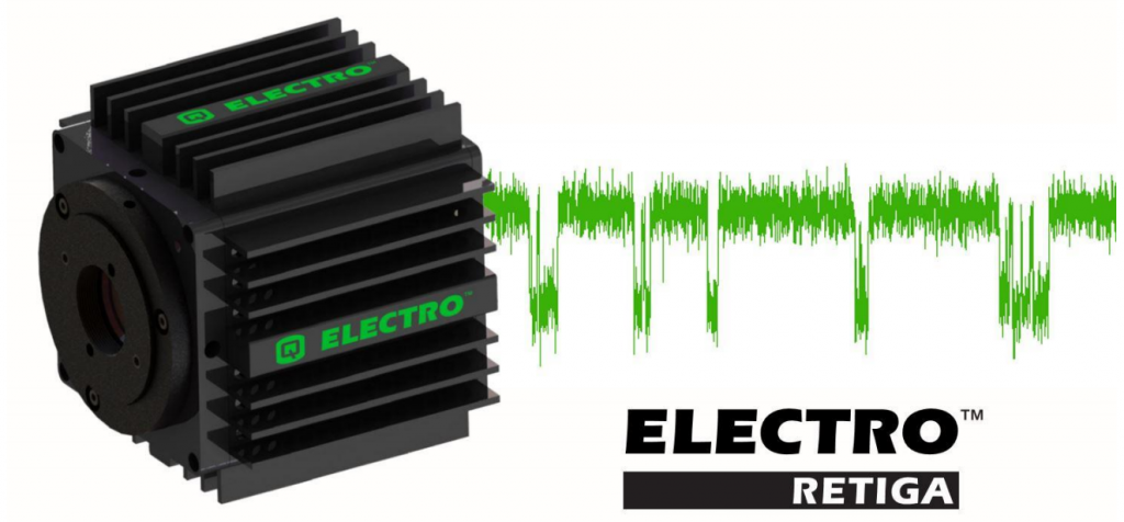
The Retiga ELECTRO
The Retiga ELECTRO was specifically designed to offer solutions for the unique challenges of electrophysiology, with high sensitivity, low noise, and vibration-free.
Specifications
Table 1: Featured specifications of the Retiga ELECTRO.
| Quantum Efficiency | 75% (at 600 nm) |
| Pixel Size | 6.45 x 6.45 µm |
| Sensor Size | 1360 x 1024 (11 mm diagonal) |
| Read Noise | <5.5 e– RMS |
| Dark Current | 0.036 e–/pixel/sec at 15 °C |
| Binning Options | 1×1, 2×2, 4×4, 6×6, 8×8, 12×12, 16×16 |
High-Quality Imaging
The Retiga ELECTRO is an ideal imaging solution for any electrophysiology setup. It has been carefully and thoughtfully designed to deliver superior performance that meets the needs of the electrophysiologist. It delivers the excellent sensitivity and noise specifications required for high contrast imaging of cells and tissues, making the manipulation of micropipettes in their contact with cell membranes and tissues significantly easier to perform. Once contact has been made between the glass pipette and sample, the Retiga ELECTRO delivers high-efficiency photoelectron conversion across the entire visible wavelength range to capture dynamic processes.
Intelligent Quantification
The Retiga ELECTRO’s use of Intelligent Quantification provides advanced real-time FPGA algorithms to deliver better image quality.
Hot pixels are pixels that repeatedly have higher backgrounds than the majority of pixels on the sensor and are a problem because they can give false readings of the signal reported by these pixels. This happens on all CCD cameras so we invented Defective Pixel Correction which corrects hot pixels to improve the signal to noise ratio over long exposures without compromising speed. Thresholds for hot and dark pixels are determined during manufacturing, and their locations stored in the camera. When Defective Pixel Correction is enabled, the signal in each defect location is replaced by the average of the pixel immediately preceding and following the defect.
High Speeds
The Retiga ELECTRO can capture images at 22 frames per second (fps) at full frame, providing high acquisition rates for electrophysiology experiments. To accommodate those who need even higher frame rates, the use of available binning options and smaller regions of interest allow even higher frame rates to capture the dynamic processes of cellular activity.
Table 2: Frame rate specifications of the Retina ELECTRO
| Array Size | Frame Rate (fps) |
| Full frame (1360 x 1024) | 22 (full), 31 (binned 2×2) |
| 1024 x 1024 | 26 |
| 512 x 512 | 42 |
| 256 x 256 | 64 |
Field of View
The Retiga ELECTRO has an 11 mm field of view, allowing for the capture of multiple events in one frame. With 1.4 million pixels over a 1360 x 1024 active pixel array, electrophysiologists will achieve good fields of view while still taking advantage of the high resolution offered by 6.45 µm pixels. Having this available field of view allows for more excitable cells present in field potential applications, and can allow for the imaging of entire grown tissues with the use of proper objectives. Additionally, the varied binning options of the camera allow for maximizing photon collection per pixel in the active array while maintaining quality images.
Superior Sensitivity
The Retiga ELECTRO has a maximum quantum efficiency (QE) of 75%, resulting in high-efficiency conversion of photons to electrons. In addition to high QE at 600 nm, the Retiga ELECTRO maintains a high QE across other commonly used wavelengths. Many cameras used for electrophysiology applications struggle with the detection of fluorophores with emission profiles at the edges of the visible electromagnetic spectrum, such as Hoechst or Fura Red. The Retiga ELECTRO maintains a high QE across both.
Table 3: QE specifications of the Retiga ELECTRO. *QE supplied by the sensor manufacturers.
| Peak | Hoechst (461 nm) | GFP (510 nm) | mCherry (610 nm) | Fura Red (650 nm) | |
| QE* | 75% | 55% | 64% | 72% | 70% |
Low Camera Noise
The Retiga ELECTRO has an exceptionally low dark current of 0.036 e-/p/s so long stare applications will not suffer in image quality as other CCD cameras do. With available exposure times up to 5 s, the image quality will not suffer due to dark current at low photoelectron counts.
Embedded image processing algorithms on the Retiga ELECTRO allow the user to correct for pixel response variations.
Low Vibration
Typical CCD cameras use fan cooling which can introduce vibration into the system and can be particularly problematic in techniques such as patch clamping. The Retiga ELECTRO is able to maintain a low dark current by using a fanless thermoelectric cooling system that cools the sensor to 0°C. The result is a camera that introduces no additional vibration to the system, meaning that patch-clamp or other electrophysiology experiments no longer need to compensate for this additional noise. In addition to the reduced vibrational noise, the Electro contains mounting screws to bolt down the camera into an existing grounding plate using 1/4”-20 thread.
Additional Features Of The Retiga ELECTRO
Hardware synchronization via transistor-transistor logic (TTL) signals allows for the seamless triggering of the camera with existing hardware. Synchronizing the camera with electrical pulse administration permits simultaneous image acquisition with triggering. Researchers can use one trigger to begin the acquisition of a stream of images (Trigger First Mode), collect a frame each time a trigger is received (Strobe Mode), or have the camera expose for the duration of the trigger signal (Bulb Mode).
Grounding the camera within the rig is of vital importance to reduce the buildup of static electricity in electrical systems. The Retiga ELECTRO contains grounding pins that fulfill this purpose.
A great camera deserves great acquisition software so access to the user-friendly imaging software package Ocular comes standard with the Retiga ELECTRO. Ocular allows users to easily capture images and movies, as well as perform many other image processing functions such as color channel merging, adjustment of color channels within the image and incorporating marker bars.
Summary
The Retiga ELECTRO is the superior choice of camera for electrophysiology because it overcomes many imaging challenges presented by imaging on electrophysiology systems. Researchers will be able to convert high percentages of photons to electrons with the cameras high quantum efficiency across the entire visible wavelength range, including the outer regions of the spectrum that are often challenging to sample. The camera fanless temperature regulation system will not introduce vibrational noise into the electrophysiology setup, reducing the need for additional vibration compensation in the system. Intelligent Quantification and Defective Pixel Correction contributes to improved signal to noise for long exposures without compromising the speed of image acquisition. The incorporated grounding pins allow for safe electrical grounding and the mounting screws provide an easy place to bolt down the camera for minimum vibration. These factors, in addition to the high-quality service expected of Photometrics, make the Retiga ELECTRO a natural addition to any electrophysiology system.