
Introduction
Since the invention of the microscope, optics have been optimized to match the performance of the human eye. The relationship between field of view (FOV), magnification and numerical aperture (NA) has changed little in the past decades, with magnification typically being given by 30-60x the numerical aperture. However, since the development of large FOV sCMOS cameras, the human eye is no longer the limiting factor it used to be. In order to push for a larger FOV while retaining high quality images, the Mesolens was engineered by Dr. Brad Amos and Prof. Gail McConnell of the University of Strathclyde.
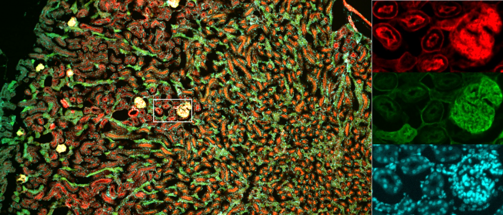
The Mesolens is a giant water-, glycerol- or oil-immersion objective that delivers 4x magnification while achieving a numerical aperture of 0.47 – enabling the capture of an unprecedented level of detail for a huge 6 mm x 6 mm FOV, with a 3 mm working distance. Compared to conventional 4x objectives with NA <0.2, the Mesolens can resolve objects over twice as small in lateral space and capture over 22 times as much light.
As a result, large tissue samples, whole organisms, or large populations of cells can be observed simultaneously with a high level of detail (Fig.1). Previously this form of imaging would have required time-consuming tiling and stitching through stage movement can be done in one shot, while the Mesolens allows dynamics to be observed for such specimens.
The Mesolens
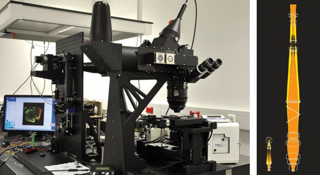
Due to its scale (indicated in comparison to a conventional objective system in the right panel of Fig.2), the objective requires entirely custom-made, high-precision lens components and is supported by a custom housing. The optics consist of 17 individual elements, up to 63 mm in diameter. An eyepiece can be added for sampling a central section of the FOV. The lens is multi-immersion, suitable for oil, water, or glycerol. Across the whole FOV, the flatness of field is <3 μm and the lens is color-corrected across the full visible spectrum.
In addition to camera-based acquisition, laser-scanning confocal microscopy can also be performed, allowing highly-resolved 3D acquisitions of an unprecedented volume. The Mesolens was also used to demonstrate a widefield, camera-based confocal technique called HiLo imaging, which can capture optically sectioned information 30 times faster than laser-scanning techniques1.
Mesolens Applications
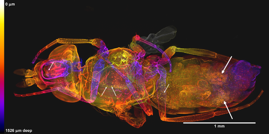
The Mesolens has many potential applications, from the screening of human genes in transgenic mice to studying large numbers of neurons in brain tissues. For example, recent Mesolens investigations of Drosophila allowed researchers to study individual organs and cells within organisms without dissection, yet still maintaining the whole organism overview, as shown by McConnell et al. (2018) and shown in Fig.3.
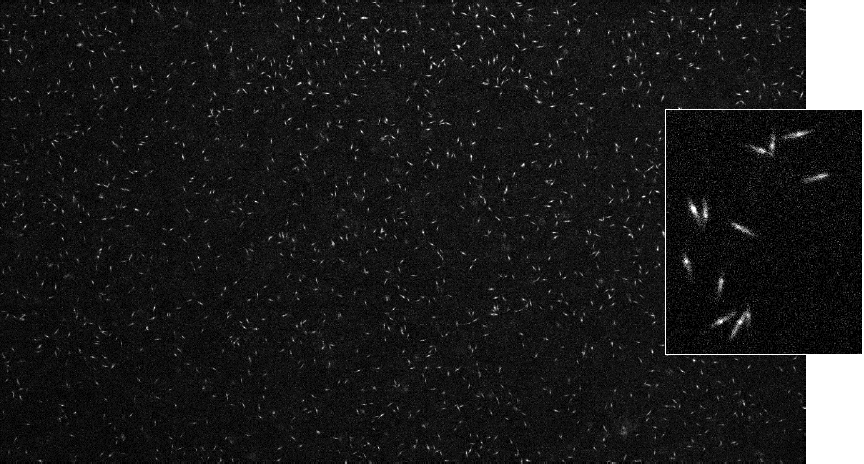
The Mesolens also has applications beyond single large organisms. Prof. Colin Brownlee of the Marine Biological Association uses the Mesolens to study interactions of large populations of plankton, imaging thousands of organisms with sub-cellular resolution and low temporal resolution necessary for calcium imaging. The image shown in Fig.4 shows unstimulated plankton, demonstrating the level of detail possible with a small-pixel sCMOS camera on the Mesolens.
Cameras for Mesolens
Due to the large FOV and high resolving power of the Mesolens, a camera with a large FOV and small pixel size is essential to take advantage of the unique capabilities of the Mesolens.
sCMOS technology features high framerates and higher sensitivity than pixel-shifting CCD or EMCCD cameras. Ideally, an sCMOS camera with small pixels and a large field of view could be used to take advantage of the unique capabilities of the Mesolens.
Summary
The Mesolens allows for high-resolution imaging over a vast FOV by making use of custom optics and a low magnification lens with a good NA. No longer limited by the human eye, the Mesolens allows for imaging of large live samples, including whole organisms.
References
- Schniete J., Franssen A., Dempster J., Bushell T., Amos W.B., McConnell G (2018). Fast Optical Sectioning for Widefield Fluorescence Mesoscopy with the Mesolens based on HiLo Microscopy. Sci Rep. 2018 Nov 2;8(1):16259. doi: 10.1038/s41598-018-34516-2.
- McConnell G. and Amos W.B. (2018) Application of the Mesolens for subcellular resolution imaging of intact larval and whole adult Drosophila. 2, s.l. : Journal of Microscopy, 2018, Vol. 270.
- Mesolens Technical Specifications. www.mesolens.com. [Online] http://www.mesolens.com/product/technical-specifications/.
- Can the Mesolens help the microbiologist? The Microbiology Society. [Online] https://microbiologysociety.org/publication/past-issues/imaging/article/can-the-mesolens-help-the-microbiologist.html.
- Brownlee C. Calcium Imaging with the Iris 15 Scientific CMOS. Marine Biological Asssociation, Photometrics Customer Reference. [Online] https://www.photometrics.com/resources/customer-references/Customer-Ref-Calcium-Imaging-University-of-Southampton