
Introduction
There are two significant challenges in biological imaging that conventional fluorescence microscopy cannot overcome:
- Biological specimens are 3D structures so to fully understand them we often need to construct 3D images
- Many processes that biologists would want to study occur inside biological structures, but other cell features such as the cell membrane block a clear view
These problems are difficult to overcome due to the need to pass light through the entire sample to illuminate the chosen image plane. This gives no control over where within the light path the returning light comes from. Light from out of focus planes, and from bright features above and below the desired plane cannot be blocked with conventional fluorescence microscopy. Confocal microscopy offers the solution to this issue.
Confocal microscopy uses optical sectioning to take multiple, thin, 2D slices of a sample to construct a 3D model from them. This process removes the out-of-focus light from other planes. Compared to other optical sectioning techniques, spinning disk confocal microscopy is high speed, high-sensitivity, and simple to implement. This makes it a very common choice for studying the 3D structure, fast dynamic processes, long-term time-lapse or details inside the cell membrane, all possible with live cells.
Spinning disk microscopy is a light rejection technique, so a high photon budget is necessary for high-quality imaging. Conventional spinning disk confocal microscopy uses a dual disk strategy to increase the amount of light reaching the sample by focusing the excitation light through microlenses on the first disk into the pinholes of the second disk. Emission light then passes back through the second disk and onto the camera. The use of two disks and microlenses is, however, expensive.
CrestOptics changes this by introducing the X-Light confocal imager: a spinning disk system with a proprietary single disk pinhole design with high light collection efficiency without microlenses to create an affordable spinning disk confocal system.
X-Light Spinning Disk
The defining feature of the X-Light system is the single disk design without microlenses. Although microlenses have improved light collection efficiency in dual disk systems, they are not without disadvantages:
- Microlenses provide no advantage for light collection, only excitation. Emitted light from the sample doesn’t pass back through the microlenses before reaching the camera.
- The microlenses f-number does not match the numerical aperture of microscope objectives or tube lens focal lengths provided by different microscope manufacturers
- With the CrestOptics proprietary disk design, the diameter of the microlenses prevents an optimum number of pinholes per unit area.
Instead of microlenses, CrestOptics have created a disk with a superior 15,000 rpm motor as well as optimizing the pinhole diameter, number of spirals on the disk, and the spiral geometrical patterns. The result is a disk with a high confocal resolution, improved out-of-focus rejection and increased signal to noise. This allows for higher light throughput through the disk, both excitation and emission, without relying on microlenses, as seen in Fig.1.
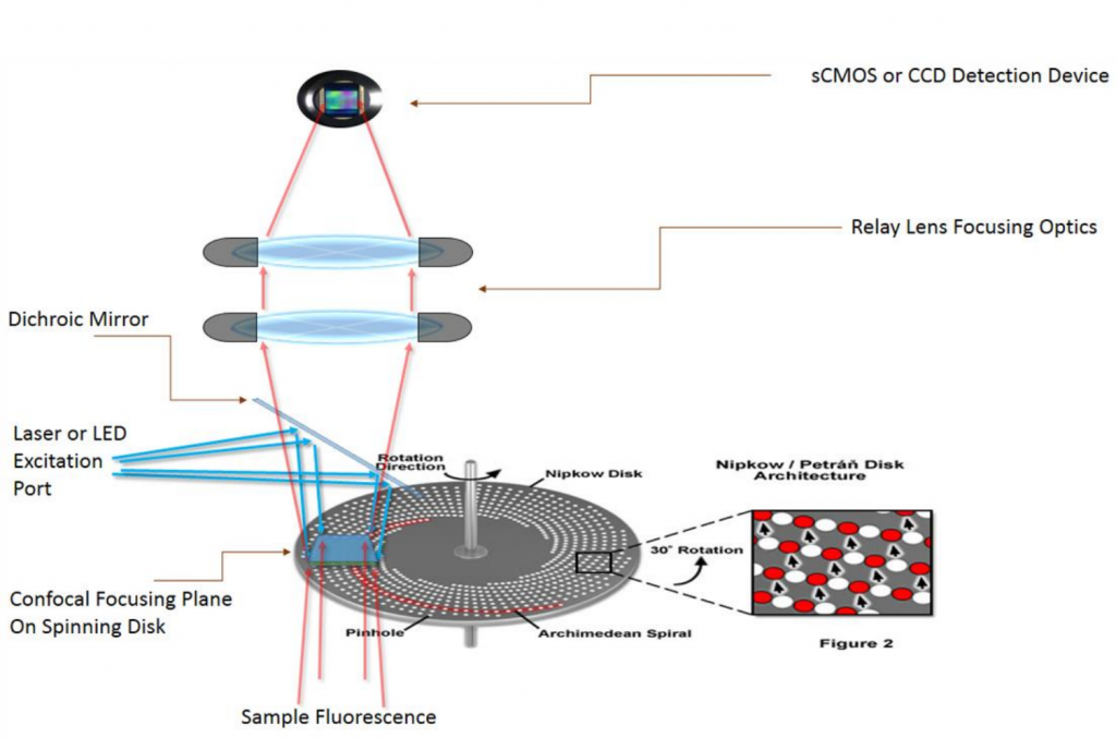
The disk includes a single or double hole pattern. With two patterns on the same disk, users can select the appropriate pinhole size for the objective lens numerical aperture or camera they are using (as seen in Fig.2). The version with larger spacing can enhance the image quality and optical sectioning ability in thicker samples thanks to the reduced crosstalk between pinholes. The disk can be easily removed and swapped with other disks, or removed entirely making the system useable as a widefield imaging system.

The X-Light also makes changes to sample illumination. Conventional spinning disk units make use of laser light sources to increase the amount of light delivered to the sample, but the X-Light has also been optimized for more affordable LED light sources. Laser light sources can still be used but LEDs have the additional benefits of a long lifespan, low energy use and produce almost no heat. This makes them much kinder to live cells, allowing them to be imaged over longer timescales. The X-Light allows for a wide variety of excitation wavelengths, multimode input and produces flat, even illumination across the entire field of view.
Large sCMOS Sensor Format
The X-Light series support larger-format camera sensors. The X-Light V2 and V3 support camera sensor sizes up to a 25 mm diagonal field of view. By taking advantage of the newest, state of the art sCMOS sensors, this allows confocal imaging of large samples at very high speeds.

Cameras For X-Light Spinning Disk
Any camera can be used with the X-Light series, but the best performance can be achieved with a sensitive camera with high speed and a large field of view.
Spinning disk confocal microscopy has historically been performed with EMCCD cameras which are highly sensitive but have a relatively small field of view and slow speed. More recently, one of the more attractive options has been back-illuminated sCMOS cameras which have equivalent sensitivity to an EMCCD camera but with the field of view, speed and pixel size advantages of sCMOS technology. This allows sensitivity and resolution to be maximized without trading off speed and field of view.
The range of pixel sizes offered by modern back-illuminated sCMOS cameras, particularly the Kinetix, can ensure Nyquist sampling with 60x magnification (with a pixel size of 6.5 µm) or Nyquist sampling with 100x magnification (with a pixel size of 11 µm). The Kinetix offers the field of view necessary to get the most out of the 25 mm diagonal of the X-Light confocal imager. Putting all of these specifications together ensures the fastest, most physiologically relevant data acquisition with the highest throughput.
Summary
The X-Light is a powerful, affordable and flexible spinning disk system that can be added to any upright or inverted microscope and combined with any steady-state light source and scientific sCMOS camera.