
Introduction
One of the most popular confocal microscopy techniques is spinning disk confocal microscopy (SDCM); a high-speed, high sensitivity technique that is reasonably simple to implement. Conventional SDCM uses a dual disk strategy that focuses excitation light through microlenses on the first disk into the pinholes of the second disk to increase acquisition speed and the amount of light reaching the sample. Emission light then passes back through the second disk and onto the camera. This method does have drawbacks, however, such as high crosstalk, low spatial resolution, limited objective lens selection, and the acquisition speed is ultimately limited by the rotational speed of the disks. For more information on SDCM, please see our full Learn page on the technique and its applications.
Swept-field confocal microscopy overcomes these limitations by using a positionable aperture plate containing a variety of pinhole columns and slit apertures instead of pinholes embedded on a spinning disk. This 1D pinhole/slit array can be swept across the sample by galvanometric and piezo-controlled mirrors to collect confocal information from structures within the plane of focus and reject out-of-focus information at high speed and high resolution with much-reduced crosstalk.
The swept-field microscope is manufactured and distributed by Bruker as the Opterra II swept-field confocal microscope.
Swept-Field Principle
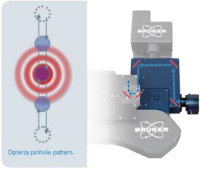
Bruker has optimized the spacing between individual pinholes (Fig. 1) which results in the system showing half or less crosstalk usually observed on a conventional spinning disk system relying on a twodimensional array (i.e. having a second disk with microlenses).
Depending on the selected microscope objective lens, the system can use three differently sized pinhole dimensions to allow the user to match the pinhole size to their imaging requirements resulting in optimal results under each condition.
In slit-scan mode, speed can be vastly increased at the expense of spatial resolution. The system has slits of four different widths to give users the choice to best accommodate their sample. The optical system is highly efficient and is able to capture more photons than spinning disk confocal microscopy with reduced back reflections from incoming light, crosstalk, and noise. The stationary pinhole apertures allow speed to be increased without the distortions typically seen at high frame rates on other scanning confocal systems.
The entire system is software controlled and the user is able to switch between pinholes/slits swiftly without the need to manually change hardware components. Moreover, a bypass mode is available to enable regular phase, DIC, brightfield, and epifluorescence on the same system. Bruker claims that the Opterra II produces a much-improved uniformity in both sample illumination and detected emission, resulting in better images and hence more quantitative data. The roll-off of the signal across the FOV is less than 10% on the Opterra II compared to 30% on an optimally adjusted conventional spinning disk system. Bruker also claims the fastest available 10-position filter-wheel (45 to 110 ms between positions), for fast emission wavelength switching. The light source is a Helios laser launch housing up to five solid-state lasers, optimized by blanking to reduce exposure time.

Cameras For Swept-Field
Various cameras have been implemented in the swept-field system but the best performance can be achieved with a sensitive camera with high speed and a large field of view. Swept-field microscopy has historically been performed with EMCCD cameras which are highly sensitive but have a relatively small field of view and slow speed.
More recently, one of the more attractive options has been back-illuminated sCMOS cameras which have equivalent sensitivity to an EMCCD camera but with the field of view, speed and pixel size advantages of sCMOS technology. This allows sensitivity and resolution to be maximized without trading off speed and field of view. The range of pixel sizes offered by modern back-illuminated sCMOS cameras can ensure Nyquist sampling with 60x magnification (with a pixel size of 6.5 µm) or Nyquist sampling with 100x magnification (with a pixel size of 11 µm). Modern back-illuminated sCMOS cameras also have the field of view necessary to get the most out of the diagonal field of view of the swept-field confocal microscope. Putting all of these specifications together ensures the fastest, most physiologically relevant data acquisition with the highest throughput.
Summary
The Opterra II swept-field confocal microscope utilizes proprietary 1D pinhole array technology in order to combine the resolution of traditional confocal systems with the speed typically associated with wide-field imaging. This allows for short acquisition times with low phototoxicity, making the technique suited for live-cell imaging.
Swept-field systems Opterra II utilizes a unique scanner design that contains both pinholes and slits, which enables flexible adjustment between speed, resolution, and intensity.
References
https://www.bruker.com/products/fluorescence-microscopes/opterra-confocal-microscopy/overview.html