
Dr. Miguel Aroso
Neuroengineering and Computational Neuroscience Lab, Institute for Research and Innovation in Health (i3s), University of Porto
Background
Dr. Miguel Aroso is a researcher involved in the functional assessment of neuronal populations using multi-electrode arrays (MEAs) and voltage imaging. This work is done in order to characterize neuronal circuits and how they communicate, how to control this activity, and how this can be used as a model for studying epilepsy and predicting seizures. The MEA electrodes can be used for recording as well as stimulation, allowing for both the recording of spontaneous and evoked activities across the network.
This, in combination with voltage imaging, allows a detailed picture of function across a network to be built up for research.
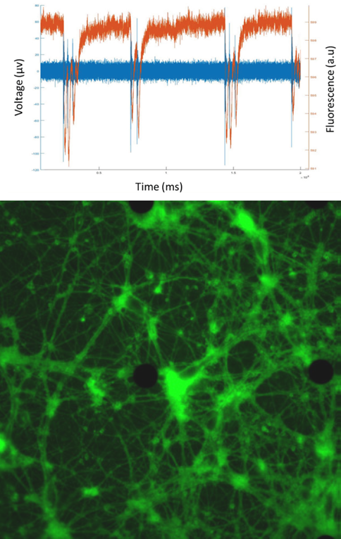
Challenge
Voltage imaging records very fast events, meaning a highly sensitive, high speed system is needed in order to capture neuronal signaling. A previous solution using a typical sCMOS camera was not able to capture this information and could not be used for voltage imaging.
Dr. Aroso outlined how speed and sensitivity were most important for voltage imaging, with an aim to image at 1000 fps or 1 kHz in order to detect relevant changes in fluorescence.
We could not record any changes in fluorescence signal with the previous system, but now we start to see voltage signals [with the Prime 95B].
Solution
The Prime 95B is an ideal solution due to the high speed and high sensitivity, allowing Dr. Aroso and the team to see voltage imaging signals for the first time. Due to the larger pixel, the Prime 95B also allows high speed imaging over a larger region of interest, resulting in more cells and MEA electrodes within the field of view.
Dr. Aroso commented on the stability of the Prime 95B signal: “we have the high speeds and now the signal is stable; we can see less than 1% change in fluorescence related with the electrical activity of the cells.” Dr. Aroso went on to state the ease of setting up the Prime 95B in MicroManager.
