Prof. Zhenyu Gao, Prof. Daan Brinks
Department of Neuroscience, Erasmus University Medical Center, The Netherlands
Background
The lab of Prof. Zhenyu Gao at the Erasmus University Medical Center studies how the brain controls motion, learning, and memory. In order to study these functions, Prof. Gao’s lab uses in vivo methods to detect electric signals in the brains of mice models. This is achieved by either utilizing electrophysiological methods or fluorescent optical methods, or a combination of both.
To visualize activity in the cortex of the mouse brain, voltage imaging can be used. This imaging technique provides an optical readout from fluorescent voltage indicators, which is an incredibly direct method of determining neuronal activities. Voltage imaging experiments are refined in the lab of Prof. Daan Brinks who collaborates tightly with Prof. Gao’s lab.

Challenge
In order to detect voltage signals in neurons, some key criteria need to be fulfilled. As imaging frequency needs to be in the range of 1-2 kHz (1000-2000 fps) in order to be able to precisely describe individual action potentials, a camera is required which is capable of this recording speed. Because of the high frame rate required, signal levels per frame will be very low, which requires a camera that has a very high sensitivity from the quantum efficiency point of view and also a low enough noise level to reliably detect even minute changes in the signal. Only by maximizing signal collection and minimizing noise contributions can a camera detect signals at low enough exposures (less than 1 ms) to operate at 1 kHz or more.
Signal levels of the currently used Archon voltage indicator reports signals from the soma (cell body) of neurons. While signal levels in electrophysiological methods can resolve even very low signal levels with high temporal resolution, voltage indicators work on the basis that their reported signals sometimes are only encoded in 1-10% increases (or decreases) in their baseline signal. These small fluctuations in signal need to be accurately collected and analyzed in order to determine neural function from voltage imaging data.
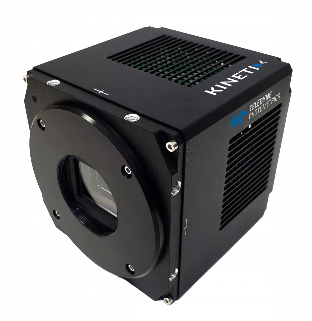
The Kinetix is the perfect solution for our requirements to detect voltage signals in vivo because it is the optimal solution – namely being fast and sensitive.
Solution
The Kinetix sCMOS presents a groundbreaking combination of both speed and sensitivity, making it a proven and ideal solution for demanding applications like voltage imaging.
The Kinetix Speed Mode images at 500 fps across the full 29 mm field of view, increasing to 1000-2000 fps at smaller regions, even to over 100,000 fps for extreme speed applications. This kind of speed is only possible thanks to the low read noise and near-perfect 95% quantum efficiency of the Kinetix.
Prof. Gao told us that “the Kinetix is the perfect solution for our requirements to detect voltage signals in vivo because it is the optimal solution – namely being fast and sensitive”. In particular, another benefit of the Kinetix is that it can image very fast and still provide a much larger field of view at the same time than previous camera solutions. This enables Prof. Gao’s lab to image many neurons at once at high speeds, putting the neuronal activities in context with each other, eventually allowing a correlation between sensory stimuli, cortical activity, and behavioral consequences.

