
Dr. Markus Gräfe
MSc. Marta Gilaberte-Basset
Optical Quantum Technologies
Fraunhofer Institute for Optics and Precision Engineering
Background
Dr. Gräfe and Ms Gilaberte-Basset’s research and development centres around quantum imaging light sources. The group is currently building a new light source that will make use of quantum imaging to permit excitation of samples in the UV range, whilst detecting in the visible range, through the manipulation of correlated photons.
The new light source offers promise for applications in cell biology due to the necessity for imaging at 400 nm in the UV coupled with the lower available QE in this range. Once developed, the new light source will permit imaging of a subject with UV light, whilst permitting detection in the 800 nm region where camera QE is much higher. The light source is based on a photon pair source, which permits the generation of correlated photon pairs. One of the photons is at 396 nm and the other is at 810 nm, one photon interacts with the object whilst the other is sent to the detector. The quantum nature of the correlated photons permits detection at the camera.
Once developed and optimized, the system will then be tested with optical setups to see how useful it may be for biological imaging in custom imaging setups.
Challenge
The light source is in the early stages of development and so Ms Gilaberte-Basset is currently testing the performance of the light source in the visible ranges before moving on to the more challenging UV wavelengths.
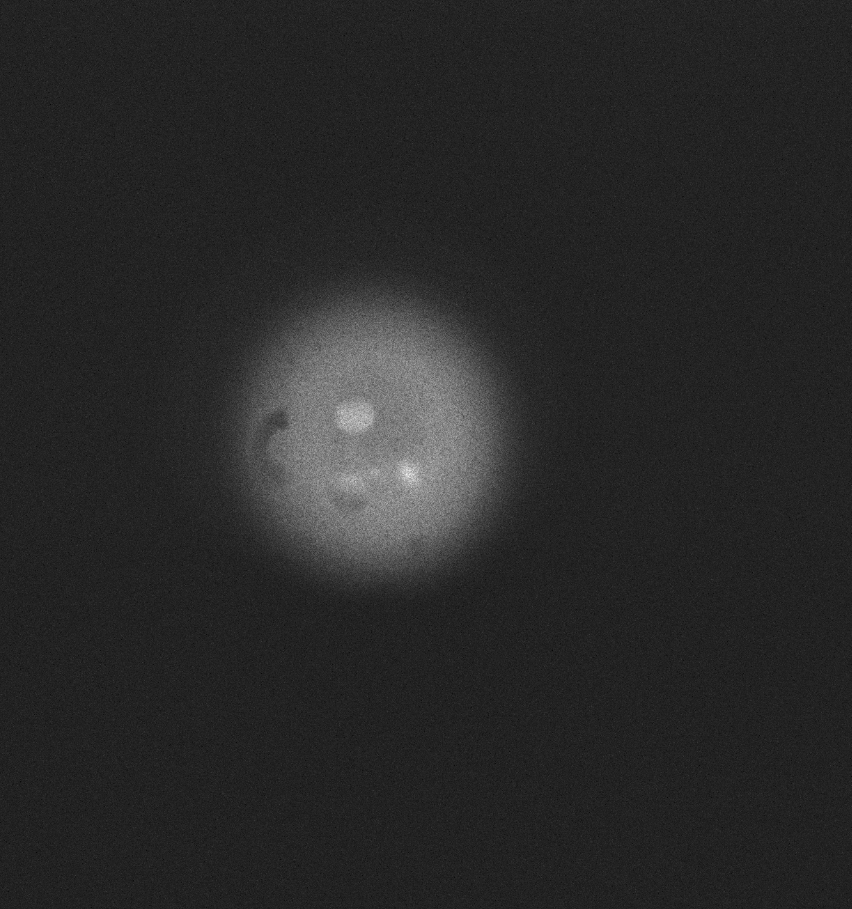
Ms Gilaberte-Basset told us: “We require a camera to check the performance of our light source. To do this, one of the samples we use consists of an opaque sheet that contains holes to allow light through, and we then assess the contrast using the camera. We have several different patterns of sample with differing hole size so that we can check how good the source is and how good is the resolution is.”
The system inherently delivers extremely low light which is essential in order to not damage the samples when illuminating with UV light, and therefore sensitivity is very important. Ms Gilaberte-Basset told us, “We were previously using an EMCCD camera to test the light source as we thought it was necessary for the sensitivity, but after checking that the imaging system is efficient enough, we replaced the EMCCD with the sCMOS which offers higher resolution due to the smaller pixel size.”
The pixel size of the Prime BSI is great, it has much smaller pixels compared to an EMCCD so it can give us much better resolution which has been a huge benefit to us. The camera itself is also much smaller which is ideal for making the system more compact
Solution
The group is now using the Teledyne Photometrics Prime BSI to check the efficiency of their light source. Ms Gilaberte-Basset shared, “We checked the system with the Prime BSI and it worked perfectly. One thing that is very important is that we want to bring the system to market. This means that if it can be more compact, robust and cost effective it is better for us.”
She went on to say: “The pixel size of the Prime BSI is great. It has much smaller pixels compared to an EMCCD so it can give us much better resolution which has been a huge benefit to us. The camera itself is also much smaller which is ideal for making the system more compact. The image we get out of the sCMOS is much better if we are checking small features, and we can get very fast acquisitions that allow us to record videos with the camera.”
