Dr. Philippe Bastin, Principle Investigator
Bastin Lab, Institut Pasteur, Paris
Background
The lab of Dr. Philippe Bastin at Institut Pasteur is primarily interested in improving understanding of the trypanosome parasites, which are significant in human health due to their role in sleeping sickness. The trypanosomes also offer a useful experimental model to increase understanding of cilia and flagella function. The lab therefore does a significant amount of cell biology investigations including live cell imaging studies, four color immunofluorescence (IF) and dSTORM imaging.

Challenge
Dr. Bastin’s lab studies protein trafficking in the flagella of trypanosomes. The challenge is that these proteins move quickly, at a rate of 2-5 microns per second in a very narrow (300 nm) environment, which is very close to the resolution limit for light microscopy.
Imaging is a tradeoff between time resolution and sensitivity, even at 100 ms exposure the particle has moved by half a micron during the exposure time leading to smearing of the trains, so it is quite challenging. When the train moves to the end of the flagella they then split into 3 smaller trains making them more difficult to detect.
Dr. Bastin was looking for a camera that had good sensitivity to decrease the exposure time. If the camera was too slow, it wouldn’t capture the movement. The previous EMCCD was used for its sensitivity, but this was limiting the resolution due to the large pixels. Four-color immunofluorescence imaging was also challenging, as when imaging dyes with longer wavelengths, such as Cy5, quantum efficiency is low.
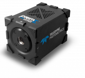
The combination of increased sensitivity and signal to noise ratio meant the 95B was the best camera choice.
Solution
Dr. Bastin is now using the Prime 95B to reduce exposure time for better time resolution while imaging protein trafficking through the flagellum. “The combination of increased sensitivity and signal to noise ratio meant the 95B was the best camera choice. For us, one of the most important things was reducing the exposure time to get better time resolution, whilst maintaining sensitivity. Even with very short exposure times of 10 ms we could start to detect the protein trains with the 95B. As the diameter of the flagella is near the resolution limit the Prime 95B with a 100X objective was the best choice for us offering a combination of speed, signal to noise ratio and sensitivity making it ideal for our applications.”
Dr. Bastin also anticipates the use of the 95B for quadruple immunofluorescence in the future. He shared, “For the long wavelengths such as Cy5, quantum yield was previously an issue. We tested the Prime 95B with this technique and we were amazed at the quantum yield of the camera, which allows us to reduce the exposure time for this type of acquisitions.”
Dr. Bastin also mentioned that he anticipated making use of the large field of view in these types of experiments. He told us, “It is advantageous for scoring cell phenotypes, such as knock out models for understanding flagella construction and motility. More cells in the field of view gives more data for each acquisition maximizing data collection and minimizing acquisition time.”

