Simon Berger, Doctoral Student
Andrew deMello, Principal Investigator and Professor for Biochemical Engineering
deMello Group, ETH Zürich (Switzerland)
Department of Chemistry and Applied Biosciences
Background
The deMello Group at ETH Zürich is engaged in a broad range of activities in the general area of microfluidics and nanoscale science. Primary specializations include the development of microfluidic devices for high-throughput biological and chemical analysis, ultra-sensitive optical detection techniques, nanofluidic reaction systems for chemical synthesis, novel methods for nanoparticle synthesis, the exploitation of semiconducting materials in diagnostic applications, the development of intelligent microfluidics and the processing of living organisms.
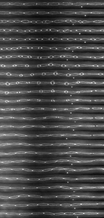
In recent years the deMello group has developed a range of microfluidic tools for the long-term imaging of living organisms, specifically the nematode Caenorhabditis elegans. Currently, work is focused on the creation of novel microfluidic devices for worm manipulation and the study of a wide range of developmental processes, previously inaccessible.
C. elegans strain courtesy of Dr. H. Ribeiro Pires and Dr. M. Boxem, Utrecht University.
Challenge
Live fluorescence imaging has seen a tremendous change over recent years. The development of sCMOS cameras has transformed image acquisition rates, fields of view and noise suppression, while also lowering unit costs. However, until recently the EMCCD has been the gold-standard for high sensitivity applications, significantly outperforming sCMOS devices with quantum efficiencies in excess of 95%, but lacking considerably with respect to sensor size and acquisition speed.
Simon Berger, doctoral student with the DeMello group, explains, “The primary challenge in live cell/organism imaging is the extraction of high quality images, both bright and with high contrast, while ensuring that phototoxicity and photobleaching are kept to a minimum. In this way, imaging does not affect sample viability and the biological processes under investigation.”
The need to ensure low photobleaching/phototoxicity often limits attainable image quality, as well as the frequency and detail with which images can be acquired. While the higher sensitivity associated with EMCCDs can remedy the effects of photobleaching/phototoxicity, the acquisition rates, the small fields of view and limited dynamic range, severely limit their usefulness for in vivo imaging.
The Prime 95B [Scientific CMOS camera] allowed us to acquire high contrast fluorescence images using low excitation intensities, and subsequently allowed us to image over longer periods of time and at higher frequencies than previously possible. This allowed for intrusion-free study of many sensitive developmental processes.
Solution
Compared to its peers, the Prime 95B Scientific CMOS combines the features of sCMOS cameras (high acquisition rates, large sensor size, low noise and high dynamic range) with the exceptional sensitivity previously only available through EMCCDs.
Simon explains, “The Prime 95B allowed us to acquire high contrast fluorescence images using low excitation intensities, and subsequently allowed us to image over longer periods of time and at higher frequencies than previously possible. This allowed for intrusion-free study of many sensitive developmental processes.”