
Prof. Zhen-Ming Pei
Department of Biology, Duke University, North Carolina, US
Background
The lab of Prof. Zhen-Ming Pei is interested in the early signalling events by which plants sense environmental signals and decode them to give the appropriate responses. Upon perception of external signals, cell surface receptors trigger an increase in cytosolic free calcium concentration, which is mediated by ion channels. Prof. Pei’s long-term goals are to identify these receptors and ion channels, isolate their interacting components, and assign molecular functions to them.
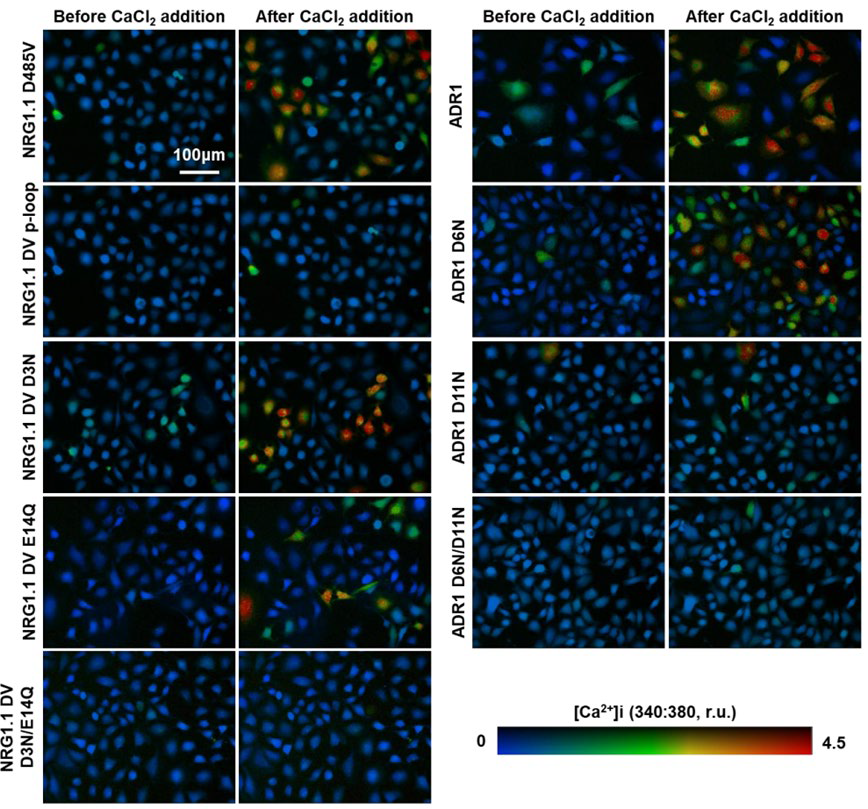
An example of Prof. Pei’s research comes from a recent Science publication, concerning the plant immune response surveillance system consisting of intracellular nucleotide–binding leucine-rich repeat receptors (NLRs) capable of triggering immunity in response to pathogen activity, leading to activation of plant defences.
The lab currently uses a multidisciplinary approach including biophysics, biochemistry, cell biology, molecular genetics, and function genomics, in order to dissect the signalling cascades of external calcium as well as nitric oxide in the model plant organism Arabidopsis.
Challenge
Calcium imaging and live-cell imaging both come with their own challenges, requiring a camera that is sensitive enough to obtain a signal while also maintaining a fast imaging rate. In order to acquire images quickly enough to observe calcium activity a short exposure time is necessary, which in turn reduces the time available to collect signal, resulting in a low signal level. Cameras for this application would need to maximize signal collection and minimize noise levels in order to get a high signal-to-noise ratio while imaging at speed.
For Prof. Pei’s calcium imaging experiments, Fura-2 fluorescence imaging was performed using a Zeiss Axiovert microscope equipped with two filter wheels and an sCMOS camera. With excitation at ~350 nm and emission at ~500 nm, another challenge is using a camera with high sensitivity at a wide range of different wavelengths of light.
The [Prime 95B] camera is very good and we have not yet pushed it to the limit, we hope to use them more in the future!
Solution
The Prime 95B camera represents the ultimate in sCMOS sensitivity, featuring a large 11 μm pixel optimized for Nyquist at high magnifications and for low signal, high sensitivity imaging. The Prime 95B also operates at up to 80 fps across the full frame, allowing for easy capture of fast calcium signals while maintaining high sensitivity and a large field of view to fit in as many cells as possible.
Prof. Pei made use of the Prime 95B in their recent Science publication, imaging HeLa cells with the Fura-2 calcium indicator. Prof. Pei gave us his opinion on the Prime 95B sCMOS, saying “The camera is very good and we have not yet pushed it to the limit, we hope to use them more in the future.”
Reference
Jacob, P., Kim, N. H., Wu, F., El-Kasmi, F., Chi, Y., Walton, W. G., Furzer, O. J., Lietzan, A. D., Sunil, S., Kempthorn, K., Redinbo, M. R., Pei, Z. M., Wan, L., & Dangl, J. L. (2021). Plant “helper” immune receptors are Ca2+-permeable nonselective cation channels. Science (New York), 373(6553), 420–425. https://doi.org/10.1126/science.abg7917
