
Dr. Christy Landes
Professor of Chemistry, Electrical & Computer Engineering, and Chemical & Biomolecular Engineering
Rice University, Texas
Background
Christy Landes’ lab at Rice University uses point spread function (PSF) engineering by phase modulation to increase the information recoverable from a two-dimensional image. Phase modulation in three-dimensional (3D) super-resolution fluorescence microscopy is achieved by mixing the phase of fluorescent light in the Fourier plane of the microscope’s detection path with a specially designed phase mask. In an alternate implementation of this geometry, the Landes group has shown that rotation of the phase mask can be used to increase the time resolution of traditional wide-field imaging experiments. The Landes lab has also designed a novel stretching lobe phase mask that can encode both temporal and 3D information in separable observables in the PSF response.
Using these approaches, they study single-molecule structure-function dynamics in chromatographic separations and in live cells. Specifically, they aim to understand early endosomal trafficking, and protein dynamics at interfaces, with spatial and temporal precisions better than otherwise available from existing optics and detectors.
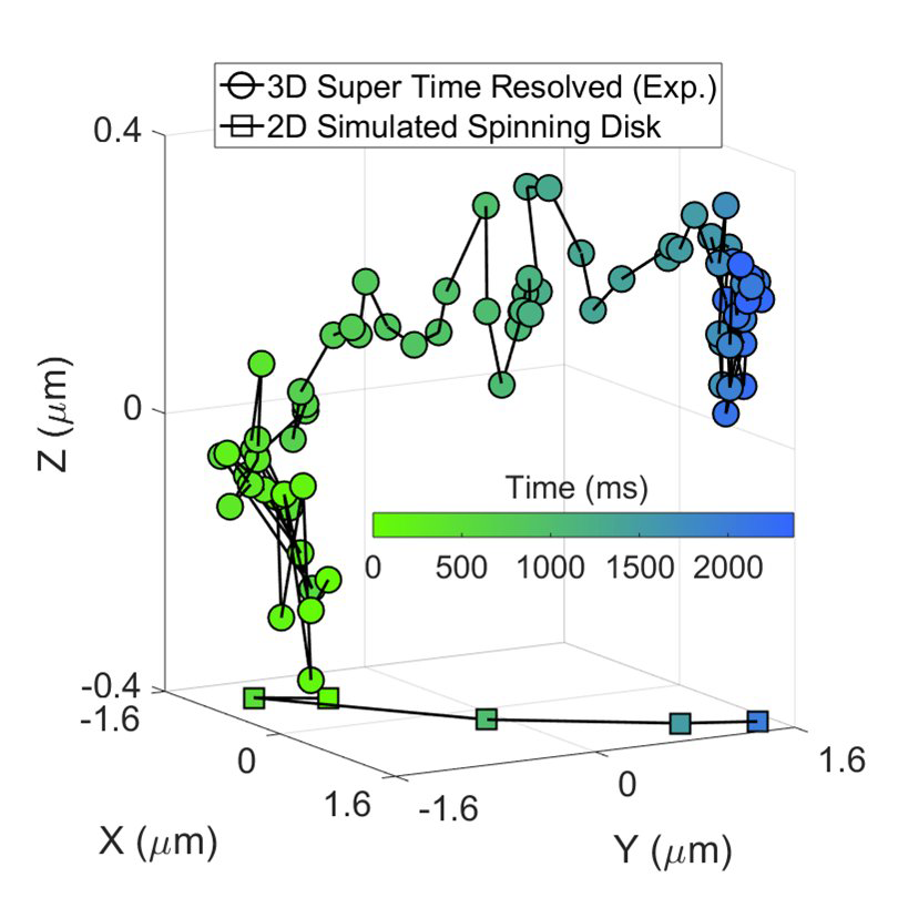
Figure courtesy: Jorge Zepeda O., graduate student in the Landes lab.
Challenge
Chayan Dutta, Post doc in the Landes group, explained their imaging challenges, “Current sensor technology cannot achieve the sensitivity and speed with which we aspire to interrogate our samples. Phase modulation of the emitted light using rotating phase mask allowed us to achieve at least 20 times faster temporal resolution. However, hardware noise and lack of sensitivity diminished our ability to discriminate these “sub-frames” as precisely as we believed possible. Using EMCCD cameras, we could amplify small signal changes and sample quickly but at the expense of the excess noise factor inherent in electronic signal amplification. CCD sensors also have limited size, so capturing the full field of view (FOV) available from the microscope was challenging.”
Dr. Landes went on to say, “We tried older sCMOS technologies but their lower quantum yield and higher pixel noise meant that we were not able to improve our 3D or temporal localization precision”.
The high quantum yield for photon detection in the Prime 95B means that now we can be confident that we can detect almost every precious photon in our low signal measurements
Solution
Dr. Landes told us, “Using the Teledyne Photometrics Prime 95B back-illuminated sCMOS camera moved us closer to our theoretical maximal information recovery. The larger FOV of CMOS chips compared to CCD recovered more of the field of view possible from the microscope. The back-illuminated sensor increased the quantum efficiency of signal capture at a lower noise level than in our EMCCD cameras. This allows for a faster frame rate with shorter integration times or a lower illumination intensity to give the signal to noise level needed to recover the extra information encoded by the PSF engineering.”
Chayan Dutta added, “The larger size of the CMOS chip of the Prime 95B also allowed us to image molecules with different excitation/emission wavelengths simultaneously on our wide-field epi-fluorescence microscope, which could be particularly useful for other biological applications.”
Dr. Landes concluded, “The high quantum yield for photon detection in the Prime 95B means that now we can be confident that we can detect almost every precious photon in our low signal measurements”.
