
Dr Siddharth Deshpande
Prof. Cees Dekker Group, Delft University of Technology
Background
Dr. Deshpande’s research focuses on the use of synthetic biology approaches to study real-time dynamics of biomolecules.
While Cees Dekker’s group deals with diverse research projects, Dr. Deshpande’s work is focused on looking at biomolecular processes in vitro by applying bottom-up approaches to improve understanding of how living cells work. Specifically, the group studies key individual components of biological relevance within a confined space, such as liposomes or droplets, which are excellent scaffolds for creating synthetic cells. The size range of these containers is in the 5-50 micrometer range.
Dr. Deshpande mainly uses high-resolution time-lapse imaging to look at the dynamics of fluorescently labeled components, such as lipids, proteins, polymers, and DNA molecules, confined within microcontainers, to understand the self-organization and emerging behavior of these molecules.
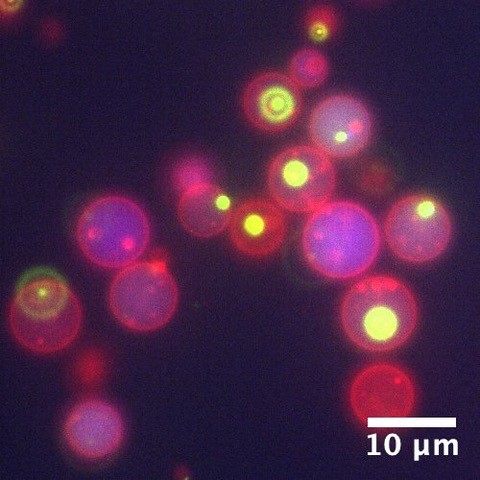
phase separated membraneless organelles (green areas)
formed within their lumen.
Challenge
Dr. Deshpande was previously using a front-illuminated sCMOS camera but wanted to improve upon the sensitivity to push the exposure time as low as possible whilst maintaining enough signal for imaging.
One challenge they faced with this type of dynamic imaging of synthetic cells was poor signal-to-noise ratio (SNR). Dr. Deshpande shared, “We struggled with SNR due to the low quantum efficiency of our previous camera and the low exposure times of 10 ms, which are necessary to prevent photo-bleaching during imaging. We want to image with the lowest exposure time possible. We also want to image at 100 or 1000 fps, so sensitivity is really important.”
Dr. Deshpande told us that “Our previous camera also had a split sensor design which leads to degradation of image quality and image artefacts.”
We previously used to image in a single plane but now we can take z-stacks through the liposome with high enough frame rates to visualize the internal dynamics
Solution
Dr. Deshpande is now using the Teledyne Photometrics Prime BSI camera with a Ti2 widefield system.
Dr. Deshpande shared, “The main reason we wanted to buy this camera was that we wanted something really sensitive that can give high frame rates. One of our colleagues had purchased the Teledyne Photometrics Prime BSI and we were impressed by the sensitivity offered by the back-illumination.”
He went on to say, “We are really happy with the camera. We previously used to image in a single plane but now we can take z-stacks through the liposome with high enough frame rates to visualize the internal dynamics. The additional quantum efficiency also allows us to use lower exposure times and not photobleach the sample.”
The camera is also being used for experiments where the high sensitivity and speed are less important but the large field of view (FOV) plays an important role.
Dr. Desphande told us, “When we are looking at slower processes we want to have the larger FOV to collect as much information as possible. This allows us to visualize multiple objects at once and get more data for better statistics. The pixel is 6.5 microns which makes the resolution ideal, this was another major factor when choosing the camera.”
