
Prof. Erwin Peterman
Dr. Andreas S. Biebricher
LaserLaB – Physics of Living Systems, Vrije Universiteit
Amsterdam, The Netherlands
Background
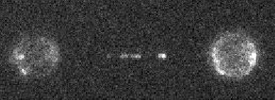
Prof. Peterman and team research protein structural and functional dynamics using combinations of optical tweezers and single molecule fluorescent microscopy. One branch of this research involves the study of proteins that bind to DNA and are involved in DNA repair and replication, in this case a human protein complex of topoisomerase IIIα, RMI1 and RMI2 that is related to DNA replication and a rare genetic disorder known as Bloom’s syndrome.
Using a custom-built inverted microscope that combines widefield fluorescence with dual-trap optical tweezers, a length of DNA (approx. 16 μm) can be stretched out between two points and imaged, observing the protein complexes bound to the length of DNA. These proteins are counted and potentially tracked using the mCherry red fluorescent protein, with an image taken every second at high magnification.
Challenge
While the advanced experimental set-up does allow for images of proteins on captured DNA to be taken, it introduces several imaging challenges. The mCherry fluorophore gives a low signal and bleaches quickly, meaning that high sensitivity and low intensity/exposure times are required.
The imaging is also limited by a 1.2 numerical aperture water immersion objective with lower light collecting efficiency, resulting in multiple lenses required to reach a 120 nm pixel size for imaging DNA and proteins.
The Prime BSI had a larger field of view than our EMCCD and was straightforward to set up and get running… we didn’t have to play around with the gain and it was good for our quantitative signal-critical applications
Solution
The Prime BSI offers top-class sensitivity with a 6.5 μm optimized pixel size to maximize light collection even at high magnification. Prof. Peterman mentioned: “imaging with the mCherry is challenging due to intense laser light being filtered out, mCherry has low signal and bleaches fast, but the Prime BSI can still see this.”
Along with high sensitivity, the Prime BSI has a large field of view allowing for more capture per frame, especially vital when imaging at low speeds for high sensitivity as fewer frames are taken and need more information.
In addition, the Prime BSI is easy to use. As mentioned by Dr Biebricher: “it was straightforward to set up [the Prime BSI] and get it running… we had to play with the gain when using an EMCCD but this was not an issue on the CMOS as they are electronically different”.
The group are using Micro-Manager with the Prime BSI, which could be set up quickly and offered several perks for this research, including QuantView to allow for easier quantification of the proteins on the DNA, and the ability to tag frames for easy analysis.
