Redmar Vlieg
John van Noort Group, Leiden Institute of Physics, Leiden University, The Netherlands.
Background
Redmar Vlieg’s research, within the group of John van Noort, primarily involves the use of two-photon microscopy to investigate biological processes in zebrafish embryos and in vitro measurements on gold nanorods (GNRs).
Due to their localized surface plasmon resonance (LSPR), GNRs have unique optical properties which allow them to be used as single-molecule sensors or very bright luminescent markers. The resonance frequency is dependent on the refractive index in the near-field of the rod, hence perturbations by small molecules can be detected by measuring this frequency shift. By exciting the GNRs via the non-linear excitation mechanism of two-photon microscopy, sensitivity can be increased to detect even smaller molecules.
Besides detection of single molecules, GNRs are used as luminescent markers. The LSPR mediates a significant increase in absorption of the excitation light as it couples with the incoming EM waves, making them as bright as quantum dots. Moreover, GNRs have the added benefit that they do not bleach and blink, and their surface can be easily functionalized for biological applications. Hence, the group investigates the merits of using GNRs as two-photon contrast agents for in vivo measurements.

Challenge
One issue that the group face when using GNRs is that when the temperature of the rods is increased the ends start to diffuse, even at relatively low temperatures. Although gold melts around 3000°C, the tips start diffusing at much lower temperatures. As gold nanorods start to lose their shape they begin to lose their aspect ratio, which causes the LSPR to shift and renders them useless for imaging.
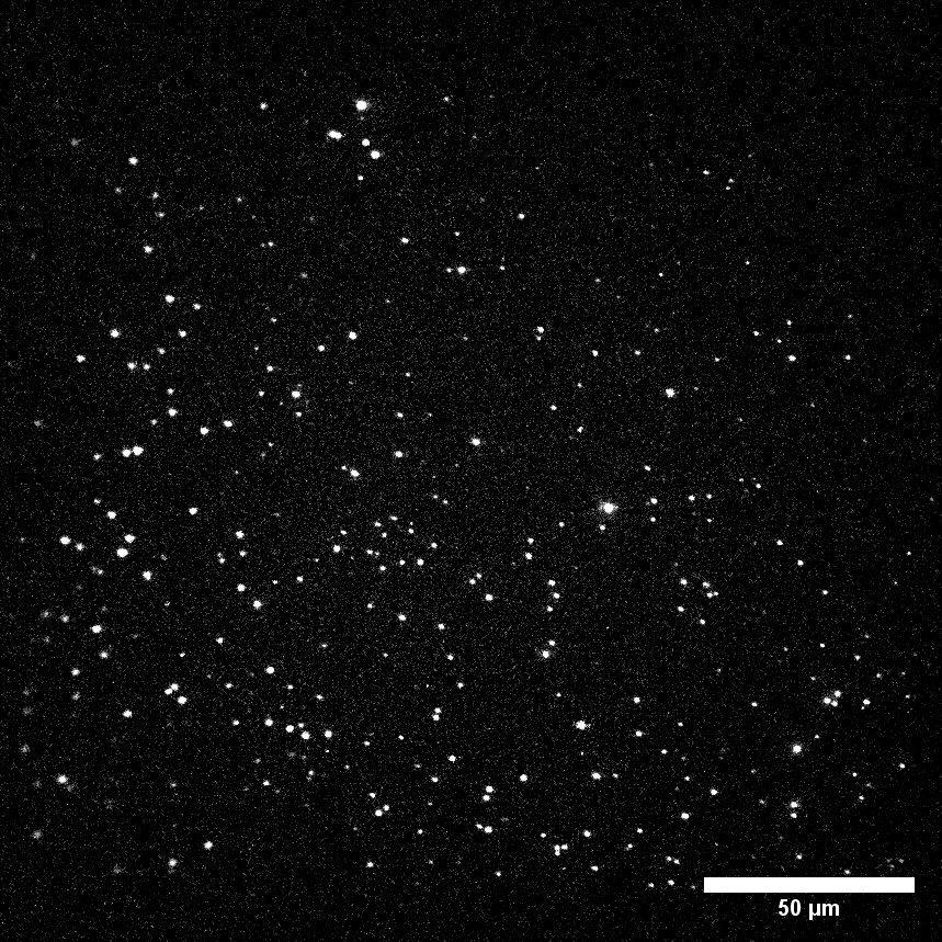
The group initially were using an EMCCD camera with a 60× TIRF oil lens for their in vitro studies. However, oil objectives have a very short working distance which is not appropriate for in vivo imaging of thick samples such as Zebrafish embryos. For this reason, they have since changed the setup to have a single 25× objective to permit a longer working distance and larger field of view, which required a more suitable camera to maximize speed and resolution.
The group therefore want to image with the lowest excitation light possible to prevent destruction of their GNR markers, as well as imaging the in vivo samples with high speed to permit single molecule tracking. For this reason, the group was interested in back-illuminated sCMOS cameras as they have high QE combined with large sensors and fast readout speeds.
The low readout noise, high quantum efficiency and large sensor size makes the Prime BSI all we need from a camera.
Solution
The van Noort Group is now using the Teledyne Photometrics Prime BSI back-illuminated sCMOS with 25× magnification on their multifocal multiphoton custom microscope, using LabVIEW to control all components of the system. Redmar Vlieg told us that “The LabVIEW drivers provided with the Prime BSI allowed us to integrate the camera exactly the way we want in our in-house build microscope.”
Redmar Vlieg went on to say, “We decided on the Prime BSI as the smaller pixel size, compared to other back-illuminated cameras on the market, made it best suitable for high resolution tracking. Moreover, our EMCCD camera, the QuantEM:512SC, was also from Photometrics, which always performed very adequately”.
Having a high QE permits the reduction of exposure time which is ideal for his applications, permitting imaging of the gold nanorods without modifying their structure. Redmar Vlieg also stated that the camera offers great benefits such as no excess noise and no gain decay.

