Dr. Peter March, Senior Experimental Officer
University of Manchester, Bioimaging Facility
Background
The research being performed at the University of Manchester has a real-world impact beyond the lab. The team is at the forefront of the search for solutions to some of the most pressing issues in biology, medicine and health. The Bioimaging Facility delivers a broad range of state-of-the-art imaging solutions to the University, Faculty of Biology, and Medicine and Health. A key technology used in biological imaging of live cells is Spinning Disk Confocal Microscopy. Spinning Disk allows for long-term, high-speed, three-dimensional imaging of live samples with multiple channels of illumination.

Sample prepared by Vicki Allen, imaged using a Yokogawa CSU-X1 with a 63x oil, 1.4NA objective.
Challenge
One of the primary reasons for using Spinning Disk Microscopy is to generate confocal images without photobleaching or damaging live samples. “Bright cells are not necessarily healthy cells,” warns Peter March, senior experimental officer at the university. “Using less GFP in cells matches their natural behavior more closely,” he adds.
Correspondingly, sensitivity is among the most important features of a camera. Until recently, and due to their ability to achieve >90% quantum efficiency, EMCCD cameras were the preferred imaging device for Spinning Disk Microscopy. March and his team typically use a 60x objective, resulting in the need to address the large pixels of an EMCCD, which require extensive optical adjustments to reach acceptable sampling levels. This severely limits field of view, making samples harder to find and capture. Additionally, EMCCD cameras cause excessive, visible noise across the sample, even at high exposures.
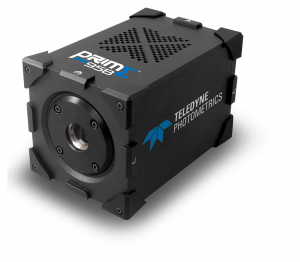
The Prime 95B [Scientific CMOS camera] is the perfect camera for Spinning Disk – the image quality is a big improvement over our EMCCDs, and the field of view makes samples much easier to find.
Solution
The Prime 95B back-illuminated Scientific CMOS is the perfect match for Spinning Disk Microscopy because it delivers much greater image quality at a higher resolution than possible with a EMCCD camera. In addition, its sensitivity is much higher than other CMOS cameras currently on the market. March is most impressed with the increase of field of view as he shares “With EMCCD cameras, finding the sample was often an issue. Field of view is all-important, and the Prime 95B is a big improvement here.”
Without the excess noise factor of EMCCDs or the pattern noise seen in 2×2 binned front-illuminated sCMOS, March says, “The difference in image quality is huge”. The Prime 95B provides the ability to capture more of a sample at equal exposure times compared to EMCCD cameras, and it produces more impressive images as a result.

