
Dr Seamus Holden
Centre for Bacterial Biology. Institute for Cell and Molecular Biosciences
Newcastle University
Background
Dr. Holden’s research lies between biophysics and microbiology, using super-resolution microscopy to study basic principles of bacterial spatial organization. In particular, the Holden Lab focuses on how the Gram-positive model bacterium Bacillus subtilis divides, and how the bacterial cytoskeleton guides the construction of a mid-cell cross-wall or septum.
The Lab is currently working to understand these biological processes using novel methods based on microfabrication, microfluidics and single molecule and super-resolution microscopy.
Challenge
Bacterial cell division takes place below the diffraction limit of microscopy, which is why the Holden Lab uses super-resolution and single molecule imaging to visualize aspects of the bacterial division process, such as the treadmilling dynamics of the bacterial cytoskeleton protein FtsZ during cell division.
The Lab’s work depends on imaging either the dynamics of cytoskeletal protein filaments using only a few fluorescent protein labels on each filament, or individual cell wall synthesis enzymes in live bacteria samples at low illumination power. This combination of low fluorophore density and low illumination intensity means that they require a scientific camera to be sensitive and have a low noise profile.
The thing that really impressed me is how uniform the sensor is – far fewer hot pixels, noisy pixels and stripes than the last generation of sCMOS cameras. I hardly see a use for EMCCDs anymore
Solution
The Holden Lab is now using the Teledyne Photometrics Prime BSI back-illuminated sCMOS to investigate how B. subtilis divide.
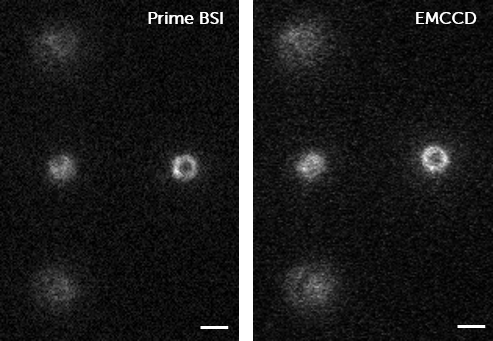
To make this decision, Dr Holden compared the Prime BSI to an EMCCD camera and was impressed with the equivalent level of sensitivity. He told us, “SNR levels are pretty indistinguishable for a tough sample of mNeonGreen-FtsZ in live cells at low illumination power.”
Dr Holden went on to say that, compared to an EMCCD, “[the Prime BSI], is faster and offers a larger field of view with 2048×2048 pixels. The thing that really impressed me is how uniform the sensor is – far fewer hot pixels, noisy pixels and stripes than the last generation of sCMOS cameras. I hardly see a use for EMCCDs anymore.”
