
Prof. Gail McConnell
Department of Physics, University of Strathclyde, Glasgow, UK
Background
The McConnell group are focused on the development of new optical microscopy methods for biomedical research and imaging.
Previously, the McConnell group were using mostly fixed cell imaging in their biological investigations, but they are now focusing on the development of techniques that will improve their live cell imaging capabilities. The group is currently working on the development of a new type of super-resolution microscopy based on the concept of standing wave microscopy.
Standing wave microscopy employs a wide field configuration that is combined with axial structured illumination to improve the resolution beyond the diffraction limit. The excitation light is composed of two beams which interfere giving rise to a standing wave, creating a sinusoidal excitation field in the axial plane. Only fluorescence molecules that coincide with the maxima of the standing wave will give rise to fluorescence.
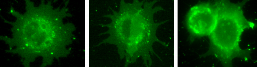
Cells plated in lbidi dishes by Gill Robb, imaged by Gill Robb and Shannan Foylan of the McConnell group.
Challenge
The McConnell group acquired a new Nikon Eclipse Ti2 widefield microscope as part of their tools and resources development project and so they started looking for a camera that would allow them to get the most out of their new system.
Professor McConnell told us, “We began to explore new options for suitable cameras to maximize the data quality from the new Ti2 instrument. We knew we wanted to use brightfield, fluorescence and DIC imaging. The microscope would also be used for live cell imaging using various magnification objectives, so we also required flexibility from the camera to be compatible with a range of objectives from 10X to 60X and compatibility with the new NIS-Elements software”.
The Prime BSI, with a 6.5 µm pixel, offers the most flexibility for use with the range of objectives we are using as well as improving the resolution.
Solution
The McConnell group is now using the Teledyne Photometrics Prime BSI on the Nikon Eclipse Ti2 widefield system. Professor McConnell shared “Following the demo of the Prime BSI, we were very happy with the camera, and have been using it every day since.”
Professor McConnell told us, “The Prime BSI, with a 6.5 µm pixel, offers the most flexibility for use with the range of objectives we are using as well as improving the resolution”.
Professor McConnell went on to say, “The customer service from Teledyne Photometrics was excellent. All our queries were responded to quickly and Teledyne Photometrics were very available and always happy to help and to offer training. I was very happy to give them our custom.”
