Dr. Shiaulou Yuan
Postdoctoral Associate in Pediatrics (Cardiology)
Yale University
Background
The Martina Brueckner lab at Yale University School of Medicine studies the genes of children with congenital heart disease. “We take a human genetics approach combined with animal models, such as mouse and zebrafish, and in vivo imaging. In particular, we do a lot of live imaging of cilia, hair-like structures on the cell surface that function as cellular antennas,” Shiaulou Yuan, a postdoctoral researcher explained. “The cilium is a tiny organelle that is packed with hundreds of distinct signaling molecules. We know these must be important because mutations in genes that are important for cilia biogenesis or signaling can cause congenital heart disease. The challenge is that for many of these genes, we simply don’t know how they can cause congenital heart disease. To understand how these genes are functioning requires us to create a whole-animal, as well as apply high-resolution live imaging approaches, that are guided by human genetics. It’s exciting because it’s the type of science that can only be done nowadays rather than twenty years ago, because of the remarkable advances in genomic and imaging technologies. It all comes together to enable us to understand the problem from new angles.”
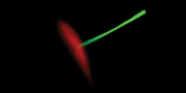
Recording by Shiaulou Yuan and Mohammed Mahamdeh using a Zeiss 63X 1.2NA water immersion objective.
Challenge
Image collection for Dr. Yuan relies not only on sensitivity and resolution, but also fast speeds. “Much of work depends on genetically encoded biosensors or GFP-knock-ins, which are endogenously tagged with a single copy of GFP. They are not that bright, but at the same time, we are also doing live imaging of mouse or zebrafish embryos that require rapid image acquisitions over several hours as the animals happily develop. We have a limited photon budget, yet we must capture a lot of images very rapidly and over a long period of time. We also need high resolution because we’re looking at tiny cilia, but also speed, because they move very fast. Finally, on top of all this, we must keep the laser excitation power low because the animal has to stay alive during all of this – the imaging needs to be gentle.”
The quantum efficiency of the camera is a really important factor for us. If we can use less excitation power, we can increase the length of our imaging and minimize photodamage to the animal. The sensitivity of the Prime 95B [Scientific CMOS camera] is truly transformative for our type of work.
Solution
“The quantum efficiency of the camera is a really important factor for us. If we can use less excitation power, we can increase the length of our imaging and minimize photodamage to the animal. The sensitivity of the Prime 95B is truly transformative for our type of work. Besides the sensitivity, the combination of speed and resolution of the Prime 95B, which is superior to an EMCCD, makes it killer for our experiments. In fact, we have been limited in the past due to insufficient camera technologies. With the Prime 95B on hand, we are now able to attack these questions.”