
Prof. Aart Verhoef
Department of Soil and Crop Sciences, College of Agriculture & Life Sciences, Texas A&M University, US
Background
The Verhoef Lab designs novel laser light sources to enhance existing and support novel imaging methods. Their systems might result in signals ranging from the detection of a few photons for super-resolution imaging, through to differentiating small differences amongst many tens of thousands of photons.
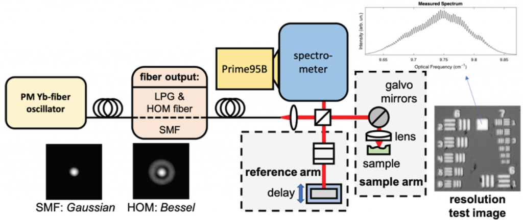
One lab focus is optical coherence tomography (OCT), which uses the ability of light to interfere with itself to map structures in tissues. Spectral OCT uses multiple colors of light to map a tissue in depth all at once.
Challenge
Spectral OCT generates a map of the position in depth, but in one shot measurement. The changes in intensity due to interference of a broad-band input source when unmixed mathematically give the same results as when moving the reference arm.
Spectral OCT uses broad-band light sources centered around 1 μm for good tissue penetration. Strong focusing of the light in the sample arm results in a high lateral resolution in the focal plane of the focusing lens, but this resolution deteriorates fast away from the focus. In order to improve the lateral resolution away from the focal plane of the OCT scan lens, the Gaussian beam illumination can be replaced by Bessel beam illumination, at the expense of optical losses.
With less power returned from regions of the sample, a camera with a large dynamic range, high sensitivity, and a large chip size is needed in a custom-built spectrometer.
Typically, a camera with only a few lines of pixels is used in such spectrometers. However, when working with Bessel beams (which have a pi phase-shift between the central peak and the surrounding weak ring), such cameras do not allow to distinguish between a phase-shift across the vertical direction of the beam and the absence of fringes.
Due to the low noise characteristics [of the Prime 95B] we can detect small objects with high fidelity even far away from the focal plane of the imaging lens… such as tiny defects in the middle of a seed.
Solution
The Prime 95B meets the requirements for a camera with a large dynamic range, large field of view, and high sensitivity.
Prof. Verhoef told us about the Prime 95B CMOS, “The Prime 95B offers a pixel size of 11 μm, that allowed us to construct a compact spectrometer matching the spatial resolution of the OCT imaging system without dramatically oversampling the resolution. The large chip size of the 95B almost exactly matched the spectral extent of our broad-band Bessel beam light source. The high frame rates that can be achieved with the 95B allow us to obtain high-resolution OCT images within a short time. The 95B allowed us to demonstrate a substantial improvement of focal depth provided by Bessel beam OCT.”
