Introduction
The key specifications to look for in a camera for calcium imaging will depend broadly on the type of calcium imaging being performed. From the perspective of the camera, the simplest way to divide calcium imaging would be into two application areas; neuronal calcium imaging and cardiac calcium imaging.

Neuronal Calcium Imaging
Introduction
Neuronal studies are used to increase our understanding of how the brain and nervous system work to better understand neurological disorders and investigate potential therapeutics. Many neuronal studies focus on the action potential which is how neurons transport an electrical signal from one to another to elicit an eventual response e.g. muscle contraction or glandular output.
In neurons, an action potential is nearly always accompanied by an influx of calcium ions so optically measuring calcium is often used as a proxy for observing action potentials. One of the distinct advantages of using a camera to image this is the ability to monitor hundreds of neurons at once and observe neural circuits.
What Is Typically Imaged
The specific biological question being asked will determine what needs to be imaged but the data obtained will broadly answer a quantitative or qualitative question.
A qualitative study may ask questions like:
- Is the cell active?
- When did the event happen?
- Is there a calcium signal?
Whereas a quantitative study may ask questions like:
- How bright is a structure relative to others?
- How much did signal intensity change over time?
- How long is the rise time or decay time?
The key difference between these studies will often be the speed required of the camera, where a qualitative study will typically have a lower speed requirement than a quantitative study.
However, calcium imaging is not fast enough to follow the full sequence of an action potential (to accurately study this, voltage imaging is the preferred technique). What this means is that the speed of the camera typically only needs to keep up with the speed of the calcium signal which will often be tracked using GECIs, such as GCaMPs or RCaMPs, and falls somewhere between 30-100 Hz.
Key Camera Requirements
High speed is important, but as discussed above more than 100 Hz is not usually necessary. Most sCMOS cameras can reach this speed.
A large field of view is also important. Firstly, to match the imaging system so the camera delivers as much as the microscope offers but more importantly to have the capability to image full neural circuits or larger samples such as live brain in vivo, large tissue slices or any cleared, fixed samples. Most microscopes allow up to a 19 mm field of view which is matched by many sCMOS cameras, but some allow up to 25 mm which is less common in scientific cameras. A home-built optical system will only be limited by the optics used which may go beyond 25 mm.
High sensitivity is also recommended for imaging at high speed with low exposure times, especially for red dyes where camera quantum efficiency often falls off. Back-illuminated sCMOS cameras with 95% quantum efficiency are recommended for this, which tend to maintain a high quantum efficiency even into the red. These cameras also typically have between 1-2 e– read noise to allow very low signal detection.
Another, more application specific, specification would be a high full well capacity which is needed when signals are expected to change rapidly from very low to very high values. Typical sCMOS camera full well capacities are in excess of 30,000 e– which is usually enough for most applications. Larger pixel sCMOS devices can have more, up to 80,000 e– if this is required.
Camera Examples
Prime BSI Express
One of the most popular cameras we have for neuronal calcium imaging is the Prime BSI Express which fits all of the key camera requirements whilst having a very simple USB interface. More information can be found on the Prime BSI Express page but at a glance this camera offers:
• 95 frames per second (full frame)
• 18.8 mm diagonal field of view
• 95% quantum efficiency
• 1.0 e– read noise
• 40,000 e– full well capacity
• 6.5 µm pixel size


Kinetix sCMOS
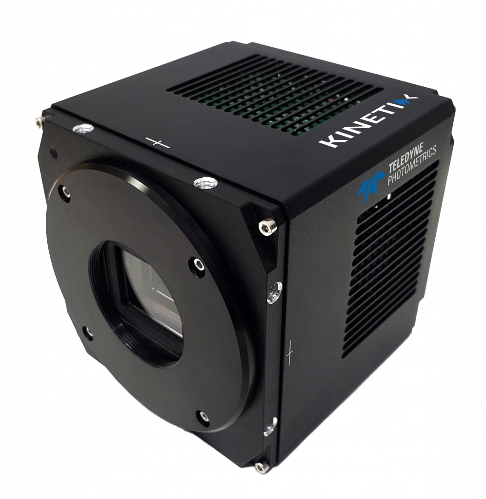
When additional speed and imaging area is required, we also offer the Kinetix sCMOS which improves on the capabilities of the Prime BSI Express. This camera is also the more popular choice when calcium and voltage imaging will both be used on the same system as the Kinetix meets the speed often required for voltage imaging applications. More information can be found on the Kinetix page but at a glance this camera offers:
- 500 frames per second (full frame)
- 29.4 mm diagonal field of view
- 95% quantum efficiency
- 1.2 e– read noise
- 6.5 µm pixel size
Cardiac Calcium Imaging
Introduction
Cardiac studies are used to investigate the electrical activity of healthy and diseased hearts, as well as the coupling between excitation and contraction in cells and tissues. This provides insight into life-threatening cardiac arrhythmias (abnormal heart rhythms) and aids design of anti-arrhythmic therapies.
In cardiac cells, the excitation-contraction coupling (ECC) process starts with the electrical excitation of the heart and ends with the contraction of the heart muscle. To enable this, the action potential opens voltage-gated Ca2+ channels in the cell membrane which increases Ca2+ concentration in the cell. Increasing Ca2+ concentration triggers further Ca2+ release from the sarcoplasmic reticulum to release even more Ca2+. This is called a calcium spark.
The summation of sparks from calcium release sites results in the calcium transient which causes contraction.
What Is Typically Imaged
Unlike in neuronal calcium imaging, the action potential is rarely studied using calcium imaging. Instead, cardiac calcium imaging typically focuses on observing calcium sparks and calcium transients.
These are both incredibly fast events with calcium sparks being 40 ms or less in duration and the sarcoplasmic Ca2+ release step of the calcium transient having a time to peak of 20-30 ms for cells paced at 2 Hz.
Cardiac calcium studies are therefore usually conducted at much higher framerates than neuronal calcium studies – it’s not uncommon to image at 500 Hz+. This also means the indicators used are different because GECI binding kinetics are too slow to operate at this speed. Instead, small molecule indicators like Indo-1 and Rhod-2 are preferred.
Key Camera Requirements
High speed is critically important, as discussed above 500 Hz+ is typical. To reach this speed, most sCMOS cameras will need to use a small region of interest which often restricts how much of the sample can be seen.
A large field of view is also important. Firstly, to match the imaging system so the camera delivers as much as the microscope offers but more importantly to have the capability to image live, whole hearts or other large samples such as tissue sections. Most microscopes allow up to a 19 mm field of view which is matched by many sCMOS cameras, but some allow up to 25 mm which is less common in scientific cameras. A home-built optical system will only be limited by the optics used which may go beyond 25 mm. As stated above however, field of view is likely to be restricted to reach the required speed.
High sensitivity is also highly recommended for imaging at such high speed with low exposure times, especially for red dyes where camera quantum efficiency often falls off. Back-illuminated sCMOS cameras with 95% quantum efficiency are recommended for this, which tend to maintain a high quantum efficiency even into the red. These cameras also typically have between 1-2 e– read noise to allow very low signal detection.
Another, more application specific, specification would be a high full well capacity which is needed when signals are expected to change rapidly from very low to very high values. Typical sCMOS camera full well capacities are in excess of 30,000 e– which is usually enough for most applications. Larger pixel sCMOS devices can have more, up to 80,000 e– if this is required.
Camera Examples
Kinetix sCMOS
One of the most popular cameras we have for cardiac calcium imaging is the Kinetix sCMOS which is our highest speed, largest field of view sCMOS camera to minimise the compromise needed to achieve the required high speeds. The increase in imaging area over a typical sCMOS camera to reach 1 kHz is nicely illustrated in Figure 5.
More information can be found on the Kinetix page but at a glance this camera offers:
- 500 frames per second (full frame)
- 29.4 mm diagonal field of view
- 95% quantum efficiency
- 1.2 e- read noise
- 6.5 µm pixel size


Summary
From a camera perspective, calcium imaging can be divided into neuronal and cardiac applications. Neuronal calcium imaging is typically slower and can usually be performed very well with a typical sCMOS device like the Prime BSI Express. Cardiac calcium imaging is typically a faster technique which often requires a small region of interest to reach the speed required. A high-speed camera such as the Kinetix sCMOS can overcome this while also being suitable for voltage imaging on the same system when multiplexing.
