
Introduction
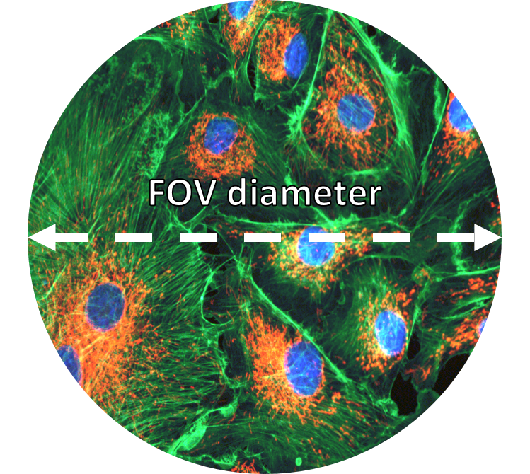
Microscope field of view (FOV) is the maximum area visible when looking through the microscope eyepiece (eyepiece FOV) or scientific camera (camera FOV), usually quoted as a diameter measurement (Figure 1). Maximizing FOV is desirable for many applications because the increased throughput results in more data collected which gives a better statistical measurement for detecting subtle effects and also decreases time needed at the microscope.
The FOV of a microscope is ultimately limited by a number of factors, such as the objective lens, the tube-diameter of the microscope’s internal optical-system, the eyepieces, the scientific camera sensor size and the camera mounting adaptor
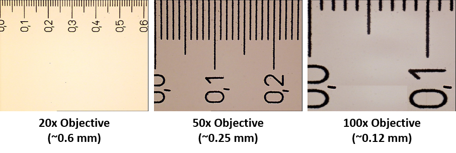
It’s usually possible to find the maximum FOV of the microscope by referring to the field number (FN) displayed on the eyepieces and on some objective lenses. The field number is simply the maximum FOV of measured as a diameter the objective or eyepiece in millimetres, so an objective lens with a field number of 18 would have a maximum FOV of 18 mm. However, the field number always assumes no magnification so to calculate the actual FOV, the field number should be divided by the objective magnification:
Field Of View = Field Number ÷ Object Magnification
A 20x objective with a field number of 18 would actually have a FOV of 0.9 mm. Likewise, a 100x objective with a field number of 18 would have a FOV of 0.18 mm. The more an object is magnified, the smaller the field of view will be. Therefore, when looking to increase FOV, one of the first considerations should always be whether it’s possible to decrease magnification (Figure 2).
Matching Scientific Camera FOV To Microscope FOV
Using the field number to calculate microscope FOV works well when imaging using the eyepieces but not when imaging using a scientific camera. Like most digital cameras, scientific cameras use square or rectangular sensors. This means that a scientific camera cannot capture the whole, circular FOV that the microscope is capable of. Instead, the camera FOV must fit inside the microscope FOV (Figure 3).
Camera specification sheets will display the camera FOV as a diagonal measurement (usually in millimeters). Ideally, the diagonal camera FOV should match the diameter of the microscope FOV to capture as much of the available image as possible. However, this does mean that the horizontal and vertical FOV of the camera will be less than the microscope diameter.
It’s possible to use a camera with a larger diagonal FOV than the microscope to capture the entire microscope FOV (Figure 4). However, this is not optimal as there will be substantial vignetting at the corners of the image. Ideally, when choosing a scientific camera, it should have a diagonal FOV that matches the specifications of the microscope it will be used with.
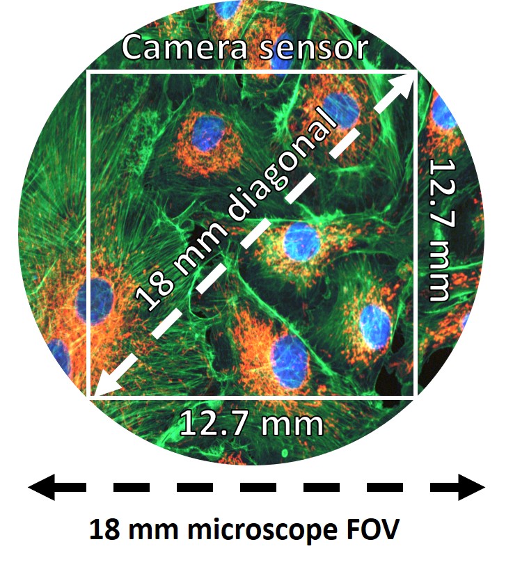
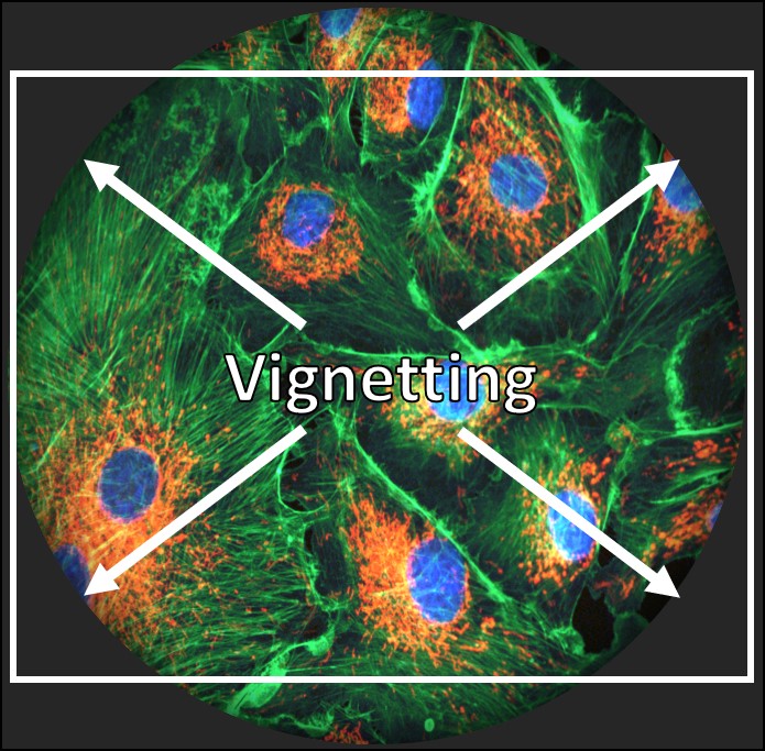
Figure 4: A camera sensor with a larger diagonal FOV than the microscope FOV would show considerable vignetting in the corners of the image.
Matching Adaptor FOV To Camera And Microscope FOV
A microscope C-mount or F-mount adaptor is needed to connect a scientific camera to the microscope camera port. The mount threading is standardized which means that a C-mount adaptor will connect to all scientific cameras that connect via C-mount. However, the adaptors are microscope specific which means that although any C-mount camera will connect to a C-mount adaptor, the adaptor will only fit microscopes of the matching brand.
Adapters can have lenses in them to magnify or demagnify the image before it reaches the camera. This can be used to better match the camera FOV to the microscope FOV. For example, if the camera has an 11 mm diagonal FOV but the microscope is capable of an 18 mm FOV, a 0.67x adaptor would demagnify the image and allow it to be displayed on the 11 mm camera. However, this increase in FOV comes at the cost of reduced resolution.
If the goal is simply to attach the camera to the microscope, a 1x adaptor contains no additional lenses and provides no additional magnification or demagnification. This is often the preferred method as it introduces no additional lenses into the system. Every extra lens reduces the number of photons reaching the camera by 3-4% so many researchers will try to avoid this.
Adaptors can also affect the microscope and camera FOV depending on the type of adaptor used. A C-mount adaptor is the most popular microscope camera adaptor and is restricted to a maximum 22 mm FOV. The F-mount adaptor is a larger format adaptor capable of reaching >30 mm FOV.
The development of larger FOV microscopes and scientific cameras that can take advantage of the F-mount is relatively recent – at the time of writing only one commercially available 25 mm microscope exists. Most modern microscopes have a 19 mm or 22 mm FOV and are therefore still able to use the C-mount. The largest format spinning disk confocal systems are also limited to a 22 mm FOV.
Choosing a Camera to Maximize Microscope FOV
At Teledyne Photometrics, we aim to create cameras that can optimally match the FOV of all modern microscopes (Table 1). For this reason, the Prime 95B Series comprises a 19 mm camera, a 22 mm camera and a 25 mm camera. Additionally, the Prime BSI and Iris 9 both fit a 19 mm microscope FOV and the Iris 15 fits a 25 mm microscope FOV. The Kinetix is our largest format sensor which is able to be used to get the maximum FOV out of any system up to 29 mm.

By recognizing that FOV requirements can be highly variable, we are able to better serve the needs of our customers and offer a broad range of camera FOV options.
Summary
The maximum field of view of the microscope is affected by the objective lens, the tube-diameter of the microscope’s internal optical-system, the eyepieces, the scientific camera sensor size, and the camera mounting adaptor. For optimal imaging performance, it’s best to match the microscope FOV to the scientific camera FOV to capture as much information as possible and avoid vignetting. Teledyne Photometrics cameras are designed to match these specifications to offer the maximum field of view possible.