Prof. Daniel Cohen, Dr. Julienne LaChance
Department of Chemical and Biological Engineering, Princeton University, USA
Background
Fluorescence reconstruction microscopy (FRM) takes in transmitted light from a biological sample and outputs a series of reconstructed fluorescence images that predict what the sample would look like were it labeled with a certain set of fluorophores. This is done using ‘deep learning’ with a convolutional neural network (e.g. U-Net) that can be trained for FRM.
The FRM approach enables many benefits including reduced phototoxicity, freeing up of fluorescence channels, simplified sample preparation, and the ability to re-process legacy data for new insights. With the increase of computational processing power year on year, FRM may become a standard tool to augment quantitative biological imaging.
However, FRM is currently complex to implement, and FRM benchmarks are abstractions that are difficult to relate to how valuable or trustworthy a reconstruction is. In this paper, Cohen et al. relate the conventional benchmarks and demonstrations to practical and familiar cell biology analyses to demonstrate how FRM should be judged in context. This research demonstrates that FRM performs well even with lower-magnification microscopy data, often collected in screening and high-content imaging. This research specifically presents results for nuclei, cell-cell junctions, and fine feature reconstruction; provides data-driven experimental design guidelines; and provides researcher-friendly code, complete sample data, and a researcher manual to enable more widespread adoption of FRM.

Imaging Solution
This paper involved testing FRM against ground truth fluorescence images, particularly in a low-magnification, high-content scenario. This requires a sensitive camera with a small pixel (to sample at Nyquist with low magnification objectives) and a large FOV in order to maximize data capture.
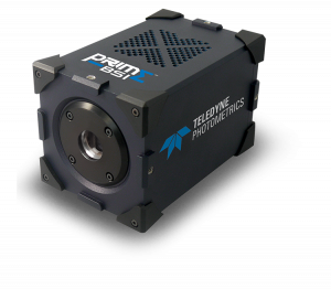
The Prime BSI used in this paper features a balanced 6.5 um pixel and a large FOV, ideal for high-resolution imaging even at low magnifications, ideal for the high-content applications for FRM testing. The 95% quantum efficiency resulted in excellent image quality and good data for processing.

