Dr. Franziska Curdt, Ms Laura Ziegenbalg
Institute for Biology and Environmental Sciences, University of Oldenburg, Germany
Background
Dr. Franziska Curdt and PhD student Laura Ziegenbalg use fluorescent microscopy techniques to investigate the magnetic senses of fish, namely the magnetic imaging of putative magnetoreceptors. To this end, Dr. Curdt has built several imaging systems to image large tissue samples, including a magnetoscope and an OpenSPIM-based light-sheet microscope.
Dr. Curdt told us about her research, “We are imaging volumes of cleared tissue in order to find out more about the magnetic sense of certain species, as the origin of this sense is still a bit enigmatic”
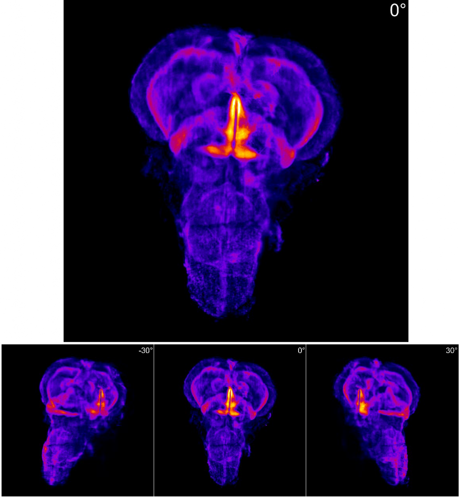
Ms. Ziegenbalg explained the choice of sample, “We want to find out how fish can perceive the Earth’s magnetic field, this involves finding brain regions that correlate with magnetic stimuli and building a reference brain atlas. While this atlas exists for zebrafish, these fish are not migratory and it is not established if they can sense the Earth’s magnetic field, so we are building a 3D atlas for the migratory rainbow trout.”
Challenge
While light-sheet microscopy with zebrafish is well established and often used as a model organism or to optimize imaging protocols, work with rainbow trout is less common. While the zebrafish brain is on the millimeter scale, the brain of the rainbow trout is many times larger, up to 2 cm. This large-format tissue imaging requires a large camera sensor in order to avoid excessive tiling and stitching.
In order to use the full aperture of the 4x objective, a small pixel is needed in order to achieve sub-cellular spatial resolution and optimize for Nyquist at low magnifications.
The small pixel size of the Prime BSI Express combined with the relatively large sensor and high speed is a big advantage.
Solution
The Prime BSI Express sCMOS features a small pixel and a large pixel array, allowing for large images to be obtained that feature high resolution at low magnifications. The high signal collection and low read noise of the Prime BSI Express maximize the signal-to-noise ratio and result in high-quality images, suitable for quantitative imaging and the construction of a 3D brain atlas.
Dr. Curdt described their experience with the Prime BSI Express, “The small pixel size of the Prime BSI Express combined with the relatively large sensor is a big advantage. We are also planning a future application involving calcium imaging. We got the Prime BSI Express with this in mind so we can do fast recording due to the high imaging speeds.”
“It was simple to get the Prime BSI Express set up in MicroManager, it worked immediately. No difficulties, no problems. I also really like the customer service of Teledyne Photometrics.”