Prof. Johann Danzl, Dr. Wiebke Jahr
Optical Imaging for Biology, Institute of Science and Technology Austria
Background
The lab of Prof. Johann Danzl focuses on research and development of cutting-edge technology to study biological phenomena at time and length scales that would be inaccessible with existing approaches. Conventional light microscopes are limited by diffraction and achieve a resolution of approximately half the wavelength of light used for imaging. At a lower resolution limit of ~200 nm (for blue light), many biologically relevant molecular details and finer morphology remain hidden.
A major focus of the Danzl Lab is the advancement of diffraction-unlimited light microscopy techniques, achieving tens of nanometer-scale resolution and better imaging in biological samples. In their interdisciplinary team of life scientists, physicists, and software engineers, the Danzl Lab tries to improve every single step of the imaging process. To this end, they are establishing novel sample preparation and labeling protocols to better mark fine structural details or highlight interactions between molecules. These samples are then imaged on custom-built microscopes offering the speed and resolution best suited for the sample at hand. At the same time, they are exploring image processing and data analysis approaches to extract more information from each sample.
Challenge
Expansion microscopy is a versatile and increasingly popular tool to acquire super-resolved images of biological specimens. Instead of improving instrument resolution, the sample is expanded to several times its original size. Fluorescent labelling of the sample can be performed before or after expansion, depending on the specific experimental requirements. Expanded samples are optically clear and the refractive index is matched to water mitigating aberrations, such that they are in principle well suited for high-resolution imaging.
Nevertheless, microscopy of expanded samples poses a number of unique challenges. Most importantly, samples are fairly large (depending on the pre-expansion size of the sample and the expansion factor). At high expansion factor and thus effective resolution, regions spanning a few cells may correspond to millimetre-sized imaging volumes. Therefore, highly parallelized microscopy methods are required to image these samples with sufficient throughput to compare different biological conditions against each other.
To image these large samples, long working distance objectives with large field numbers are preferable, but their limited NA decreases the captured signal intensity. Due to the dilution of the fluorescent markers in the expanded volume, signal intensities are decreased further.
In the Prime BSI, we found the perfect camera offering excellent signal-to-noise ratio and a quantum efficiency previously only achieved by EMCCD cameras.
Solution
The Danzl Lab built a custom light sheet microscope to image expanded samples, but needed a fast and sensitive detector to match its performance. They decided to use sCMOS camera technology due to their large chip size and high acquisition speeds when compared to EMCCDs.
The Prime BSI provided an ideal solution to the Danzl Lab for expansion microscopy imaging, offering excellent signal-to-noise ratio and a quantum efficiency previously only achieved by EMCCD cameras.
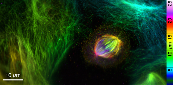
With the Prime BSI, members of the Danzl Lab could acquire expanded samples in tens of seconds to a few minutes – e.g. the entire volume around the dividing cell, corresponding to a post-expansion volume of 330 x 168 x 100 um3 (2.1×10⁹) voxels, was captured in under two minutes.