Prof. Christof Gebhardt, Mr. Devin Assenheimer
Institute of Biophysics, Ulm University, Germany
Background
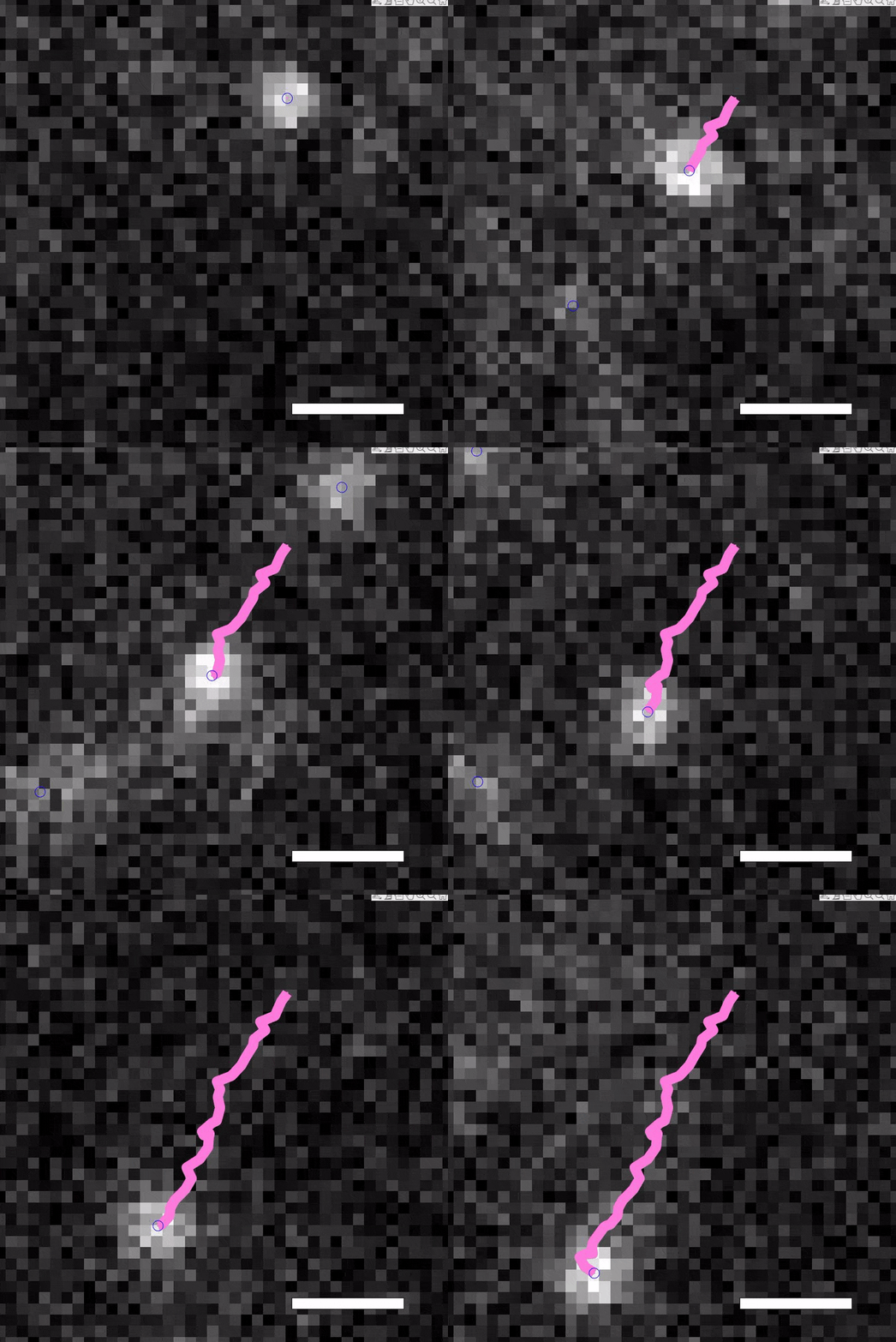
Prof. Christof Gebhardt, along with PhD student Devin Assenheimer and the team from Ulm University told us about their research. “We perform single-molecule fluorescence microscopy both in vitro and in vivo within cells, with the aim to also image small organisms. We use a light sheet-like illumination scheme where the sheet thickness can be set using a pinhole. This, in combination with organic dye fluorescent labels, gives us sufficient signal to noise for single-molecule detection even at very high temporal resolutions. We can basically image almost anywhere in a cell, we often image a couple of microns above the glass surface.”
“With live cell single-molecule experiments, we can measure the kinetics of biological molecules. For example, we can look at molecular motors and investigate how they move and measure their velocities. Conditions used in vitro with purified proteins create an artificial environment, so velocities measured in vitro might be different than what is happening in vivo in the live cell.”

Challenge
Traditional single-molecule fluorescence microscopy of fixed cells carries its own challenges, using live cells introduces additional complexity. Prof Gebhardt explained further, “Live cells cannot be permeabilized and cleared to minimize autofluorescence. Thus the background is higher compared to imaging in fixed cells.”
“To image single molecules, we need the spatial resolution high enough to distribute the fluorescent signal over a couple of pixels on the camera sensor. Therefore the magnification is such that we only have one cell in the field of view.”
“Another challenge is that live cells do not tolerate high laser power. To measure molecular kinetics, we typically want to go to a high temporal resolution of 100 Hz, so 10 ms exposure. Since we cannot increase the laser power above a critical value, this means the signal of a single molecule is only a few hundred photons. Thus, we are interested in a low read noise in order to get a good signal-to-noise ratio.
”This application requires a robust and flexible imaging device that can image with high sensitivity without losing spatial or temporal resolution, all at a high speed and across a large field of view.
One of the reasons we went for the Prime BSI was because it is fast with a large field of view, along with the low noise levels.
Solution
The Prime BSI sCMOS is the ideal solution for this application, with low overall noise and a large camera sensor with a small pixel size. Prof. Gebhardt described his experiment with the Prime BSI, “Before, we were working with EMCCD cameras due to the high signal to noise, but now we feel that the sCMOS takes over in that respect. We can also benefit from higher temporal resolution even in a low-light application. We can make the regions of interest small and go for a very high speed.”
“The big advantage of these sCMOS cameras is the high framerate possible with low signals. One of the reasons we got the Prime BSI was because it is fast with a large field of view, along with the low noise levels.”
Mr. Assenheimer spoke in terms of software and ease of use, “We are using the Prime BSI with MicroManager, the experience with the camera is good, really easy to implement. I’ve worked with it for a while now and not experienced problems.”
The Prime BSI sCMOS also allows for future improvements of experiments, with the large field of view and advanced hardware triggering systems allowing for simultaneous multifocal or multichannel imaging when paired with a splitter.

