Dr. Andreas Hans
Spectroscopy/Physics with Synchrotron Radiation Group, Experimental Physics, University of Kassel, Germany
Background
Dr. Andreas Hans is the subgroup leader of the Spectroscopy and Physics with Synchrotron Radiation group within Experimental Physics. This group is performing studies on x-ray interactions with samples and the effects on a molecular level, using x-rays to irradiate biomolecules in a vacuum and detecting the resulting emission of photons, electrons, and other subatomic particles.
As Dr. Hans outlined: “We want to mimic what happens to real biological material after exposure to x-rays, but on a molecular level… we want to see the molecular mechanisms that cause the damage to the organism, such as sunburn or cancer.”
Challenge
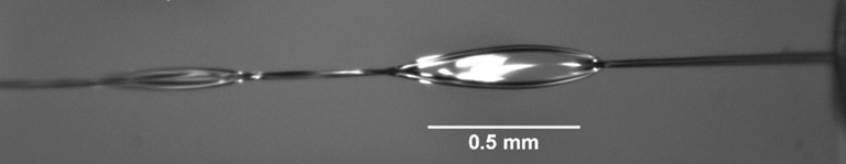
Dr. Hans wishes to expand the repertoire of samples to include liquid samples: “The problem is bringing liquid samples into a vacuum… there is a technique that injects a high-pressure beam of liquid through a pinhole into the vacuum. A tiny beam of liquid forms in the vacuum and is very hard to characterize and stabilize.” This beam is tens of microns across, meaning a sensitive camera is needed to observe and measure the water beam. In addition, two water beams can be crossed to form a very thin sheet (order of nanometres) in the vacuum. These beams and sheets of water need to be imaged with a highly sensitive and high-resolution camera for effective characterization.
In addition, there are challenges with detection, as the irradiated samples produce very low signals (such as single photons). For detection, Dr. Hans uses an amplifying device: “It transfers the photons to electrons and amplifies it to a charge cloud, which hits a fluorescing screen”. The fluorescence from this screen has a very short lifetime and needs to be detected by a sensitive, high-speed camera
Whenever a particle impinges on our amplifier we get a very short tiny flash, and due to the good contrast and sensitivity [of the Prime BSI], we can see these individual light spots and flashes
Solution
The Prime BSI presents a highly sensitive, fast, and high-resolution imaging solution for this application, both for monitoring the properties of water beams and combined sheets and for detecting fluorescent events from the amplifier.
About the water beams, Dr. Hans said: “The water sheet is very thin, in the order of nanometres, and with the camera, we can very nicely see how this behaves.” As for detecting events from the amplifier, Dr. Hans mentioned: “Whenever a particle impinges on our detector we get a very short tiny flash, and due to the good contrast and sensitivity [of the Prime BSI], we can see these individual light spots and flashes.” Overall across the different applications, Dr. Hans was pleased with the performance of the Prime BSI: “I see that it works perfectly for what we are doing and what we plan to do further, the [Prime BSI] is robust and we can get all the information we want to…it is the best one for the combination of these two different applications.”
In addition, Dr. Hans and the team also have plans to implement the Prime BSI in a custom Python data acquisition system, which benefits from the excellent software support for Photometrics cameras.